
Age-related macular degeneration (AMD) affects the macula and can progressively reduce sharp central vision. We provide comprehensive assessment, monitoring, and treatment tailored to the type and stage of AMD, with the aim of preserving vision and maintaining everyday visual function.
AMD is a condition that affects the macula, the central portion of the retina responsible for detailed vision. The macula is responsible for tasks such as reading, recognising faces, and seeing detail clearly.
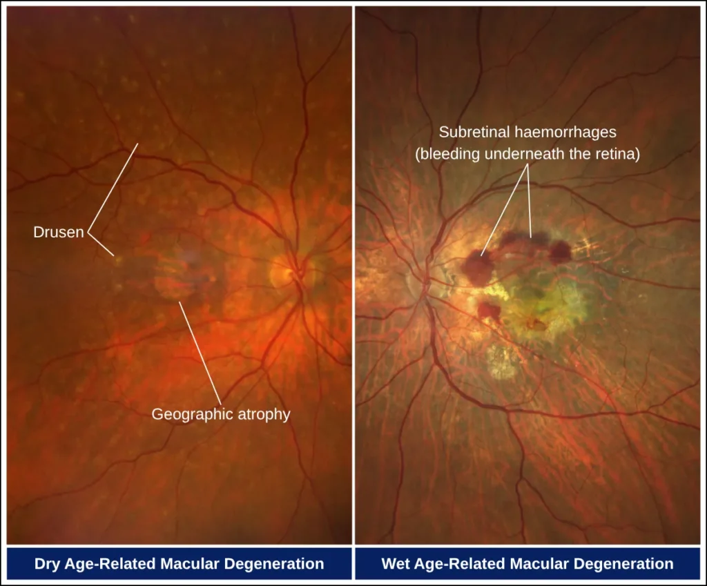
Dry AMD is more common and usually progresses gradually as the macula and its supporting tissues deteriorate. In advanced cases, areas of retinal tissue may become permanently damaged, a stage known as geographic atrophy.
Wet AMD occurs when abnormal blood vessels develop beneath the retina and leak fluid or blood. Although less common, it can cause more rapid and significant changes in central vision. Dry AMD may sometimes progress to wet AMD.

AMD develops due to a combination of age-related, environmental, and vascular factors that influence retinal health over time. Common contributing factors include:
In AMD, age-related degenerative changes develop within the supporting layers and the overlying macula, disrupting the normal structure and function of the cells.
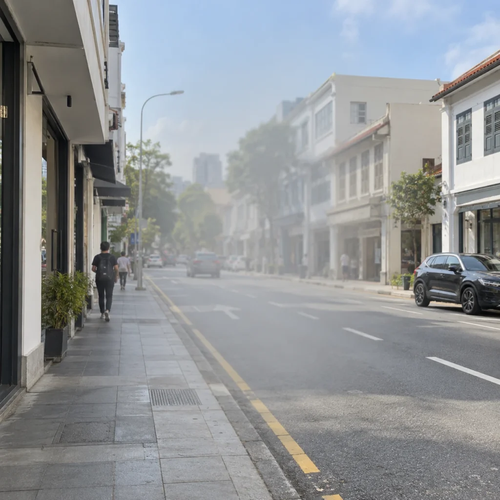
Individuals with AMD might experience symptoms such as:




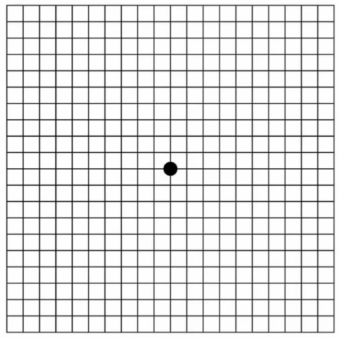
The Amsler grid is a simple way to check for changes in central vision that may occur with AMD.
How to use the grid:
Noticing any changes in your vision? Book an appointment with us for a comprehensive eye assessment.
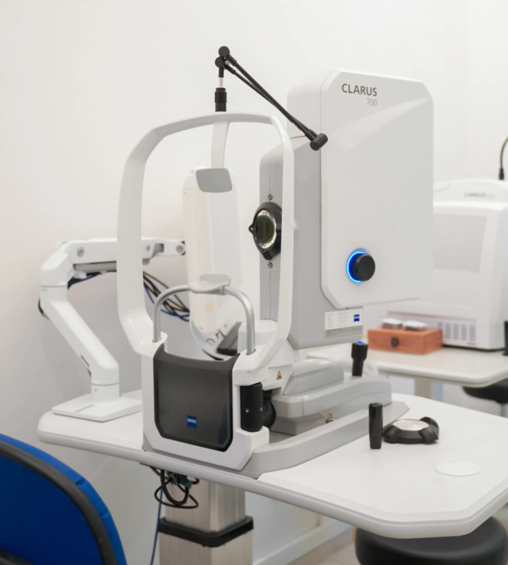
We use a combination of detailed eye examination and advanced retinal imaging to assess the macula, retina, optic nerve, and retinal blood vessels. These tests allow us to identify structural and functional changes, determine the type and severity of the condition, and establish a clear baseline for ongoing monitoring and treatment planning.
Measures how clearly you can see and helps identify any reduction in central vision.
Captures detailed images of the retina and retinal blood vessels to identify abnormalities and signs of age-related macular degeneration.
Maps areas of retinal pigment epithelium and photoreceptor loss, helping identify and monitor geographic atrophy in dry AMD. It is particularly useful for documenting changes in atrophic areas over time, including during treatment with anti-complement injections.
Uses fluorescein dye and a series of retinal photographs to assess blood flow and leakage in the macula, helping identify abnormal new blood vessels and areas of active wet AMD.
Provides cross-sectional images of the retina to assess the macula and individual retinal layers for swelling, fluid or other structural changes.
Provides a non-invasive view of the macular circulation to identify abnormal choroidal neovascularisation (CNV) and assess the abnormal blood vessel network associated with wet age-related macular degeneration.
Treatment for retinal conditions is guided by the type of condition, its severity and how it changes over time. Regular follow-up allows us to monitor the retina closely and determine when treatment is needed. When intervention is required, we will recommend the most appropriate treatment at the right time, with the aim of controlling the condition, protecting the retina and preserving vision.
AREDS2 supplements can be taken daily to support macular health and provide key nutrients involved in maintaining retinal function. The formula contains lutein and zeaxanthin, antioxidants concentrated in the macula, together with vitamins C and E, zinc and copper.
For people with intermediate AMD, regular AREDS2 supplementation has also been shown to reduce the risk of progression to advanced AMD. The supplements are taken orally as tablets or capsules, and we will advise on the recommended formulation and dosage based on your retinal findings.

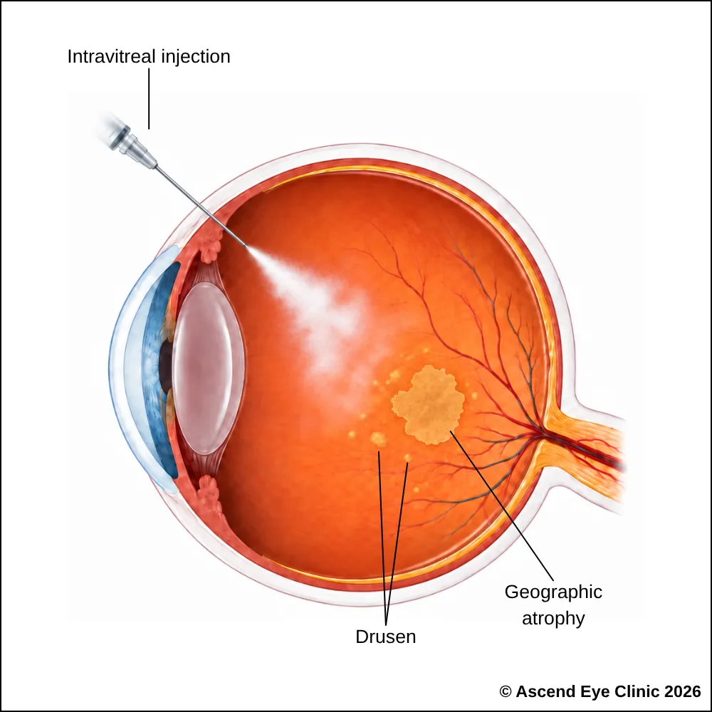
Anti-complement injections are used for geographic atrophy, an advanced form of dry age-related macular degeneration (AMD). They work by reducing overactivity of the complement system, which contributes to ongoing damage to retinal cells. Treatment helps slow the enlargement of geographic atrophy and preserve the remaining retinal tissue and vision for longer.
An intravitreal injection is performed as a quick outpatient procedure with careful preparation to keep the eye comfortable throughout. Anaesthetic drops are used to thoroughly numb the eye before the injection. Although the idea of an injection into the eye can sound daunting, the injection itself takes only a few seconds, much like a vaccination procedure, and most people feel only very mild discomfort.
Several injections are often given at regular intervals over several months to bring the retinal condition under control and achieve stability. Active treatment can be stopped and maintenance treatment can be followed with continually monitoring the retina closely with imaging during follow-up visits.
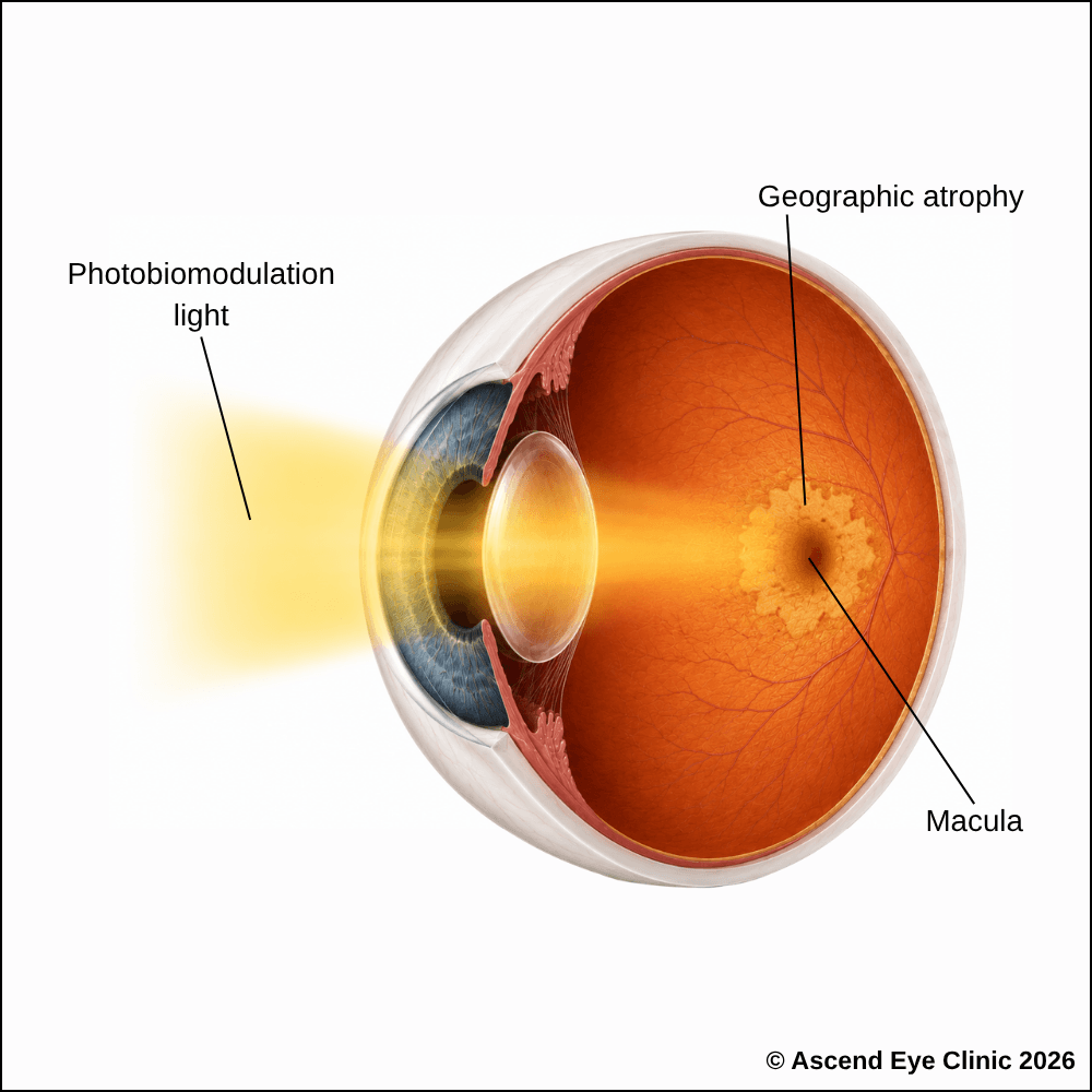
Low-level light therapy is a non-invasive treatment for dry AMD that uses specific wavelengths of light to stimulate retinal cells and support their function. The treatment is designed to support macular health, maintain vision and improve visual function in selected patients.
During treatment, you will sit comfortably while a specialised light-delivery device is placed in front of your eyes. Controlled wavelengths of light are then delivered to the retina in a series of short treatment cycles. No injection or anaesthetic is required, and each session takes only a short time.
Treatment is usually performed as a course of repeated sessions over several weeks. The aim is to support retinal function and help preserve visual function as part of the ongoing management of dry AMD.
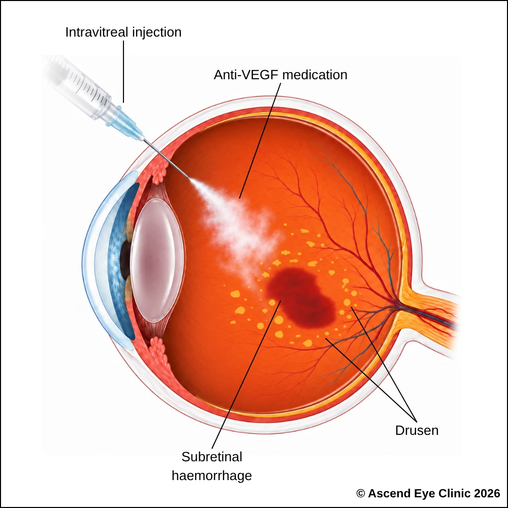
Anti-VEGF injections are used to treat wet age-related macular degeneration (AMD), where abnormal new blood vessels grow beneath the retina and leak fluid or blood into the macula. By blocking VEGF, treatment helps reduce leakage and swelling, control abnormal blood vessel growth and preserve central vision.
At Ascend Eye Clinic, anti-VEGF medications available include Eylea®, Lucentis® and Vabysmo®. We will advise on the most appropriate medication based on the appearance of the macula, disease activity and response to treatment.
An intravitreal injection is performed as a quick outpatient procedure with careful preparation to keep the eye comfortable throughout. Anaesthetic drops are used to thoroughly numb the eye before the injection. Although the idea of an injection into the eye can sound daunting, the injection itself takes only a few seconds, much like a vaccination procedure, and most people feel only very mild discomfort.
Several injections are often given at regular intervals over several months to bring the retinal condition under control and achieve stability. Active monthly treatment can be converted to maintenance treatment at a lower dosing frequency, with continued close monitoring of the retina with diagnostic retinal imaging during follow-up visits.
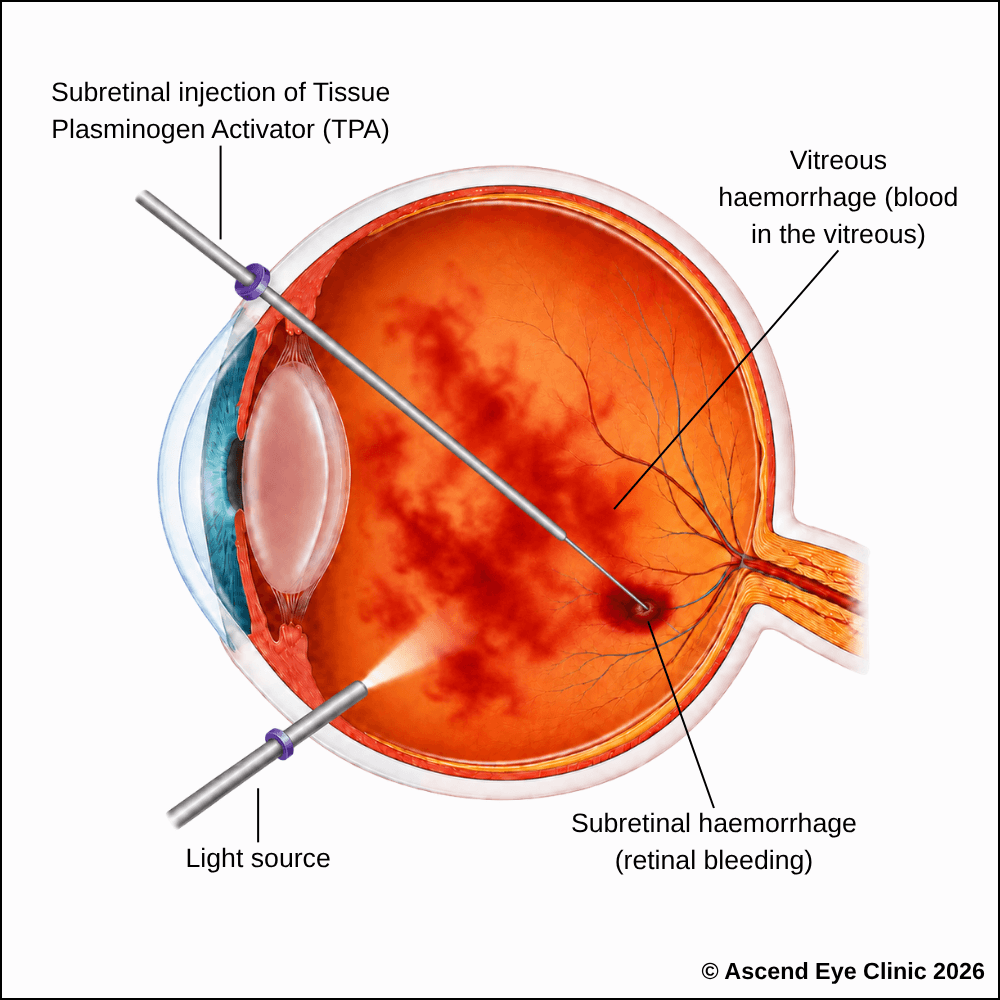
In age-related macular degeneration, abnormal blood vessels beneath the macula can occasionally cause a large collection of blood to build up beneath the central retina. When the bleeding is recent and extensive, a vitrectomy combined with a subretinal injection of Tissue Plasminogen Activator (TPA) can be performed to help dissolve the blood clot and displace it away from the centre of the macula. This reduces the amount of blood lying beneath the central retina and helps limit further damage to the macula.
Vitrectomy is a keyhole surgery performed in an operating theatre under sedation or general anaesthesia. Fine instruments are introduced through tiny ports in the eye to remove the vitreous gel. A small amount of tPA is then injected beneath the retina into the area of haemorrhage to help break down the clot. A gas bubble is placed inside the eye to help move the liquefied blood away from the centre of the macula. Anti-VEGF treatment is also used where required to control the abnormal blood vessels responsible for the bleeding.
Vision is blurred initially after surgery and gradually improves as the gas bubble dissolves and the blood clears. The extent of visual recovery depends on the size and duration of the haemorrhage and the degree of damage already present in the macula from age-related macular degeneration.
Medical Director & Senior Consultant Ophthalmologist
Ascend Eye Clinic, Singapore
From your initial consultation to treatment and follow-up care, our team will guide you through every stage of your journey. We will take the time to explain your condition, answer your questions and help you feel informed and supported throughout your care.

Your consultation begins with a detailed assessment of your vision and eye health. Depending on your condition, this may include measurements of your vision and eye pressure, examination of the front and back of the eye, and advanced diagnostic imaging.
Our team will guide you through each test and explain what is being assessed. Your results will then be carefully reviewed to understand your individual needs and determine whether monitoring or treatment may be recommended.
Every eye and every patient is different. Your treatment plan will be tailored according to your examination findings, visual needs, general health and lifestyle.
We will explain the recommended treatment, its expected benefits, possible risks and any suitable alternatives in a clear and straightforward manner. You will have time to ask questions and consider your options before deciding how you would like to proceed.
Before your treatment, our team will guide you through the necessary preparations, paperwork and medication instructions. Where applicable, we will explain how to use your prescribed eye drops and how to care for your eyes afterwards.
We understand that undergoing a procedure can feel unfamiliar or worrying. Our team will be available to answer your questions and ensure that you know what to expect before the day of your treatment.
On the day of your procedure, you will arrive at the hospital or surgical centre where your operation has been scheduled. The surgical team will help you prepare and explain what will happen at each stage.
Anaesthetic eye drops or other forms of anaesthesia may be used, depending on the procedure, to keep you comfortable. Your doctor and the surgical team will support and reassure you throughout the operation.
Most eye surgeries are performed as day procedures, although the duration and recovery process will depend on the treatment you receive. Your vision may be temporarily blurred afterwards, so you should arrange for someone to accompany you home and should not drive unless your doctor has advised that it is safe to do so.
We will provide clear instructions on how to care for your eyes, use any prescribed medication and manage your activities during recovery. We will also explain which symptoms are expected and when you should contact the clinic for further advice.
Follow-up appointments allow us to monitor your vision, assess your recovery and identify any concerns at an early stage. Our team remains accessible should you have any questions or concerns throughout your recovery.
Early assessment and timely treatment can help preserve vision and reduce the risk of further damage from age-related macular degeneration. Book a consultation at Ascend Eye Clinic for a detailed retinal assessment and personalised treatment plan.
AMD often affects both eyes, although the severity and rate of progression can differ between them. If AMD is already present in one eye, the other eye has a higher risk of developing AMD, so regular monitoring of both eyes remains important.
Yes. Dry AMD can progress to wet AMD when abnormal blood vessels develop beneath the retina and begin leaking fluid or blood. Sudden blurring, distortion or a new dark area in the central vision requires prompt assessment.
AMD progresses at different rates. Some people remain stable for years, while others develop more significant changes over a shorter period. Regular retinal examination and imaging allow us to monitor for progression.
Signs include increasing central blur, straight lines appearing wavy, difficulty reading or recognising faces, and new dark or missing areas in the centre of vision. Any sudden change in central vision requires prompt assessment.
AMD primarily affects central vision rather than the entire field of vision. Even in advanced AMD, peripheral or side vision is generally preserved. Significant central vision loss can still affect reading, recognising faces and driving.
There is currently no cure that completely reverses AMD. Treatment focuses on slowing progression, controlling active disease and protecting as much central vision as possible.
Treatment and regular monitoring help reduce the risk of further progression. Wet AMD is treated to control abnormal blood vessel growth and leakage, while dry AMD is managed with monitoring, appropriate nutritional supplements for suitable stages and control of modifiable risk factors.
The review schedule depends on the stage of AMD and whether there are signs of active wet AMD. We will determine the appropriate follow-up interval based on the retinal examination, imaging findings and any changes in vision.
Yes. An Amsler grid is a simple way to monitor central vision between appointments. Check one eye at a time and look for new waviness, blurred areas, dark patches or missing sections of the grid.
Arrange an eye assessment promptly. These symptoms can indicate the development or reactivation of wet AMD, where early treatment is important for protecting the macula.
Dry AMD involves gradual degeneration of the macula and is commonly associated with deposits called drusen. Wet AMD develops when abnormal blood vessels grow beneath the retina and leak fluid or blood, resulting in a faster and more significant change in central vision.
Dry AMD is primarily managed with regular monitoring and management of risk factors. For certain stages of AMD, nutritional supplements are recommended to reduce the risk of further progression. We will advise whether supplementation is appropriate based on the retinal findings.
Wet AMD is mainly treated with anti-VEGF medication delivered directly into the eye through an intravitreal injection. The medication suppresses abnormal blood vessel growth, reduces leakage and controls swelling within the macula.
Injections are generally given at closer intervals at the beginning of treatment. Once the retina becomes stable, the interval between injections is adjusted according to the response seen on retinal imaging.
There is no fixed number of injections for wet AMD. Treatment is tailored according to how the retina responds and whether abnormal blood vessel activity remains controlled. Retinal imaging helps guide the ongoing treatment schedule.
The surface of the eye is thoroughly numbed with anaesthetic drops before treatment. A very fine needle is then used to deliver the medication, and the injection itself takes only a few seconds. Most people experience brief pressure or mild discomfort rather than pain.
Improved or stable vision often means the treatment is successfully controlling the abnormal blood vessels. Stopping treatment while the condition remains active allows leakage or bleeding to return and can cause further damage to the macula. We adjust treatment according to the retinal findings rather than vision alone.
Treatment aims to control abnormal blood vessel activity, reduce retinal fluid and prevent further damage to the macula. The amount of vision recovered depends on how much retinal damage was already present before treatment and whether scarring has developed.
For suitable stages of AMD, AREDS2 supplements contain a specific combination of antioxidants and nutrients, including lutein and zeaxanthin, that help reduce the risk of further progression. We offer MacuBio, an AREDS2-based supplement, and will advise whether it is appropriate based on the retinal findings.
Yes. Cataract surgery can still be performed when AMD is present. Removing the cataract improves the portion of blurred vision caused by the cloudy lens, while the final level of vision also depends on the condition of the macula.
Hi, I would like to book a consultation with Dr Errol Chan from Ascend Eye Clinic
