
Polypoidal choroidal vasculopathy (PCV) can cause recurrent bleeding and fluid leakage beneath the retina, leading to blurred or distorted central vision. Accurate diagnosis and timely treatment are important to control disease activity, reduce further bleeding and maintain visual function.
Polypoidal choroidal vasculopathy (PCV) is a particular form of neovascular, or wet, age-related macular degeneration (AMD). Like wet AMD, it involves abnormal blood vessels developing beneath the retina. In PCV, these vessels form a branching network with characteristic rounded swellings known as polyps, which can leak fluid or bleed and affect central vision.
PCV is associated with changes in the blood vessels and circulation within the choroid, the vascular layer beneath the retina. Detailed retinal imaging helps us identify these abnormal vessels and the changes they cause beneath the retina, distinguish PCV from other forms of wet AMD, and guide treatment.
Increasing age, inherited susceptibility, smoking, and high blood pressure are associated with a higher risk of PCV. The condition is also more commonly seen in Asian populations.
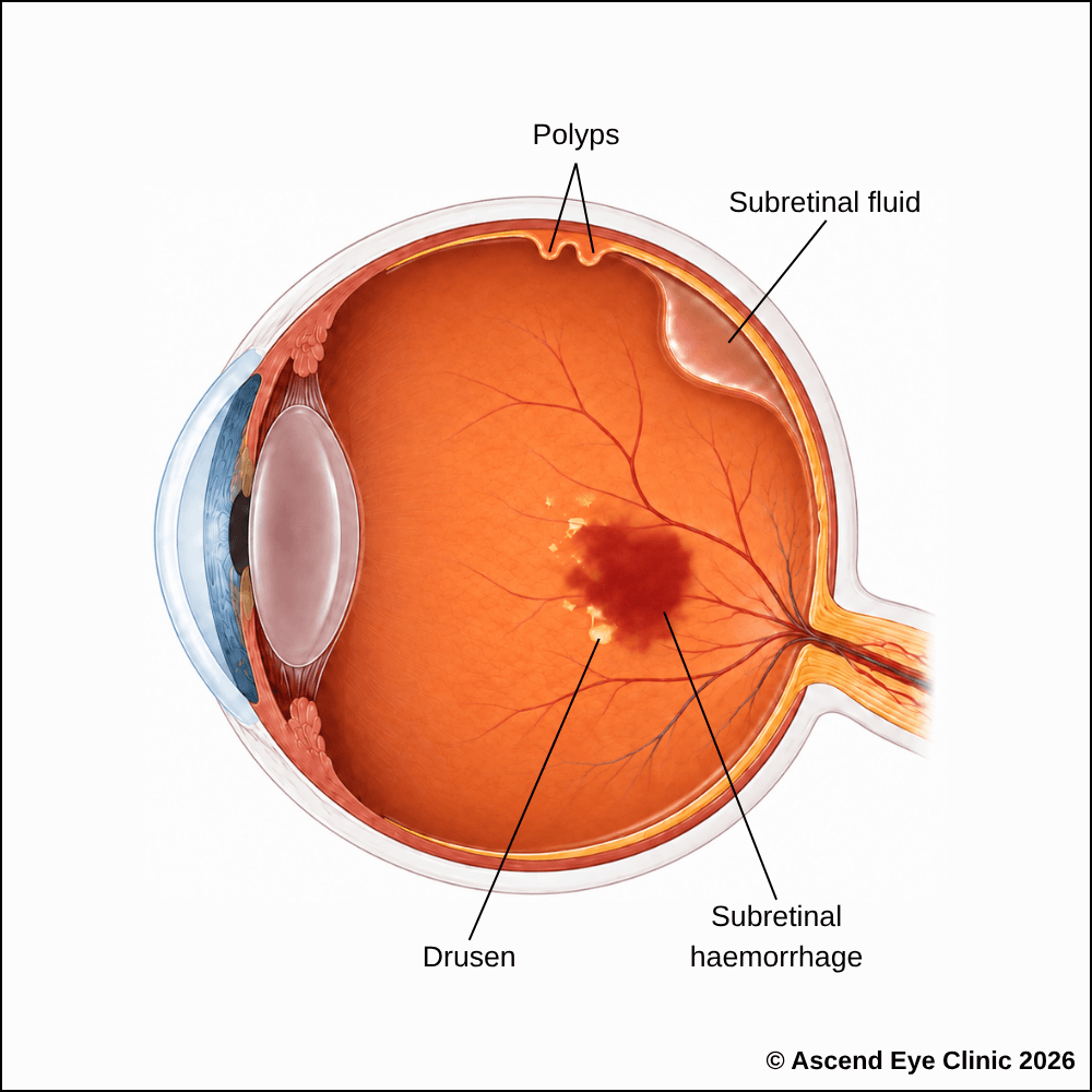


Polypoidal choroidal vasculopathy can remain quiet without causing any noticeable symptoms. This means a person can have polyps at the back of the eye without realising it. If these abnormal vessels start to leak or bleed, vision can deteriorate quickly or gradually over time, and the damage can be permanent.
When the abnormal vessels beneath the macula begin to leak or bleed, they can affect central vision, causing blurred or distorted vision, difficulty reading or recognising faces, a dark or missing area in the centre of vision, or sudden severe vision loss following significant bleeding.
We use a combination of detailed eye examination and advanced retinal imaging to assess the macula, retina, optic nerve, and retinal blood vessels. These tests allow us to identify structural and functional changes, determine the type and severity of the condition, and establish a clear baseline for ongoing monitoring and treatment planning.
Measures how clearly you can see and helps identify any reduction in central vision.
Captures detailed images of the retina and retinal blood vessels to identify abnormalities and signs of polypoidal choroidal vasculopathy.
Uses fluorescein dye and a series of retinal photographs to assess leakage and identify abnormal blood vessels associated with certain types of pigment epithelial detachment.
Provides detailed imaging of the choroidal circulation to identify underlying abnormal vessels or polypoidal lesions that may be causing the pigment epithelial detachment.
Provides cross-sectional images of the retina to identify and measure pigment epithelial detachments and assess for associated fluid or other macular changes.
Provides detailed images of the blood vessels beneath the retina to identify abnormal vascular networks associated with PCV, helping assess the extent of the disease.
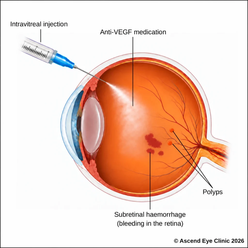
Treatment aims to control leakage or bleeding from the abnormal blood vessels, reduce fluid affecting the macula and protect central vision. The treatment plan is guided by retinal imaging, the location and activity of the lesions, and the extent of bleeding.
Anti-VEGF injections are used for diabetic retinopathy when retinal blood vessels become leaky, as in diabetic macular oedema, or when abnormal new blood vessels develop, as in proliferative diabetic retinopathy. By blocking VEGF, treatment helps reduce leakage and swelling, causing abnormal blood vessels to regress.
At Ascend Eye Clinic, anti-VEGF medications available include Eylea®, Lucentis®, and Vabysmo®. We will advise on the most appropriate medication based on your condition and treatment needs.
An intravitreal injection is performed as a quick outpatient procedure with careful preparation to keep the eye comfortable throughout. Anaesthetic drops are used to thoroughly numb the eye before the injection. Although the idea of an injection into the eye can sound daunting, the injection itself takes only a few seconds, much like a vaccination procedure, and most people feel only very mild discomfort.
Several injections are often given at regular intervals over several months to bring the retinal condition under control and achieve stability. Active treatment can be stopped and maintainence treatment can be followed with continually monitoring the retina closely with imaging during follow-up visits.
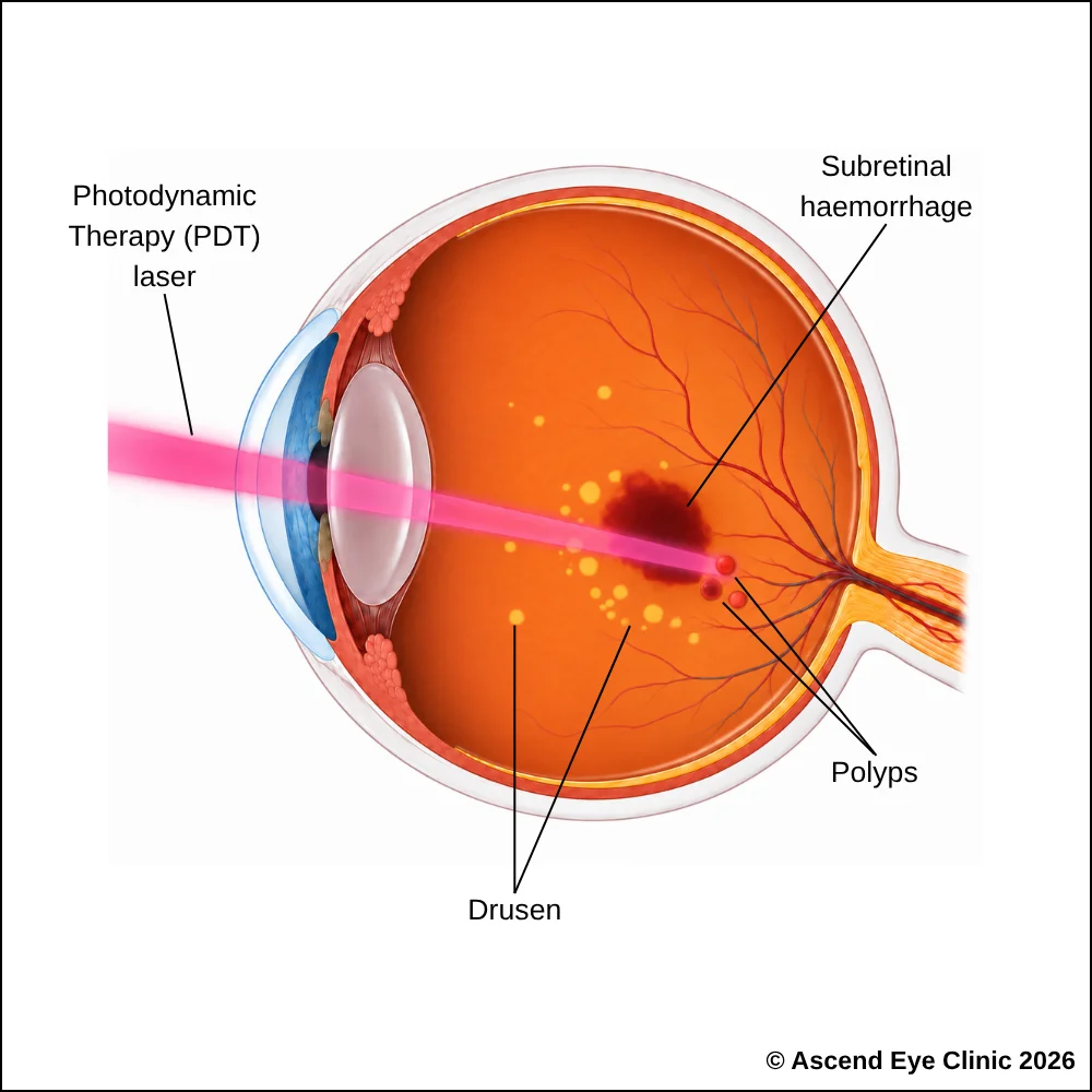
Photodynamic therapy (PDT) is a targeted laser treatment used in selected cases of polypoidal choroidal vasculopathy, particularly when prominent polypoidal lesions are causing persistent leakage or bleeding beneath the retina.
PDT is performed as an outpatient procedure and does not require a hospital stay. Treatment begins with an intravenous injection of verteporfin, a light-sensitive medication that circulates through the bloodstream and concentrates within the abnormal choroidal blood vessels. A specialised low-energy laser is then directed precisely at the area of PCV. The laser activates the verteporfin, producing a local reaction that closes the abnormal vessels and polypoidal lesions while limiting damage to the surrounding retina.
By closing these abnormal vessels, PDT helps reduce further leakage, bleeding and fluid accumulation beneath or within the retina. This allows the retina to settle and helps preserve or stabilise central vision.
PDT is frequently combined with anti-VEGF injections. Anti-VEGF treatment controls leakage and retinal swelling, while PDT provides more direct treatment of the underlying polypoidal vessels. This combined approach can be particularly useful when PCV remains active despite injections alone.
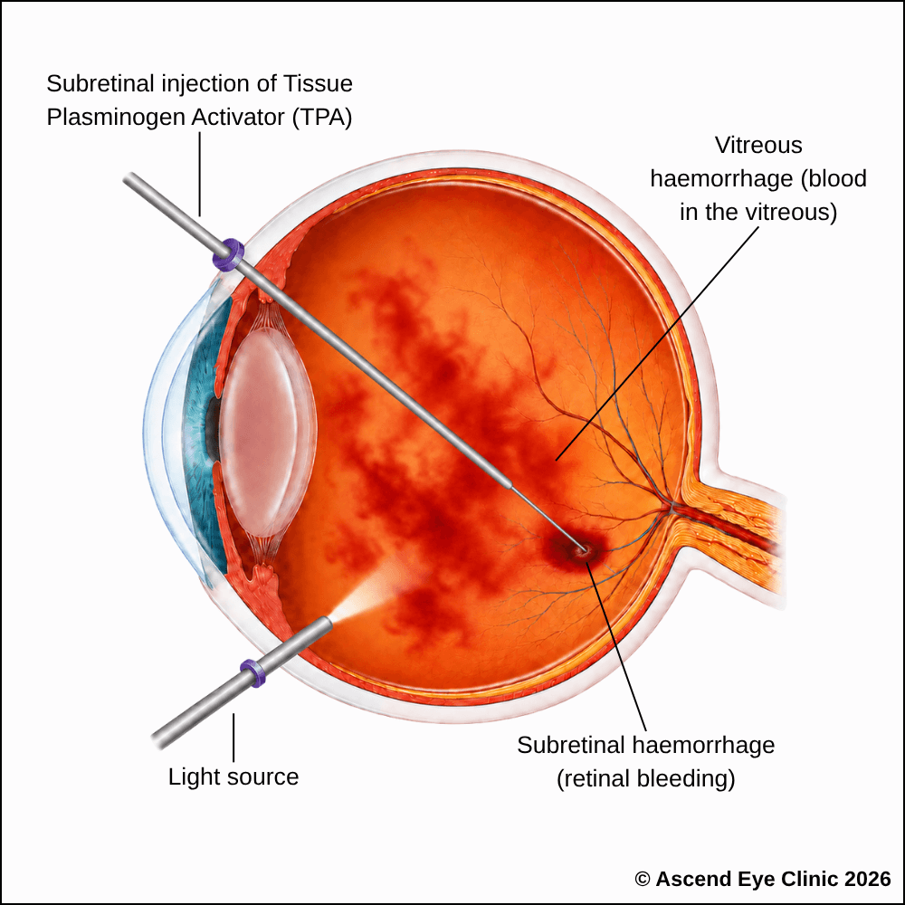
A vitrectomy allows the vitreous gel and any blood obstructing the visual axis to be removed, while also providing access to treat or displace blood affecting the central retina. In cases where there is fresh and large bleeding in the retina, an effective surgical option whereby a vitrectomy is performed together with an injection of Tissue Plasminogen Activator into the clot can help dissolve the clot and move it away from the centre of the retina, thereby recovering vision in what would have otherwise been an absolute loss of vision in the eye.
Vitrectomy is a keyhole surgery performed in an operating theatre under sedation or general anaesthesia. Fine instruments are introduced through tiny ports on the surface of the eye to remove the vitreous gel and clear any vitreous haemorrhage. A gas bubble is then placed inside the eye, which gradually dissolves on its own over time.
Vision is blurred initially after surgery and gradually improves as the eye heals and the blood clears. The extent of visual recovery depends on the amount of bleeding and how much the macula and underlying retina have already been affected by PCV.
From your initial consultation to treatment and follow-up care, our team will guide you through every stage of your journey. We will take the time to explain your condition, answer your questions and help you feel informed and supported throughout your care.

Your consultation begins with a detailed assessment of your vision and eye health. Depending on your condition, this may include measurements of your vision and eye pressure, examination of the front and back of the eye, and advanced diagnostic imaging.
Our team will guide you through each test and explain what is being assessed. Your results will then be carefully reviewed to understand your individual needs and determine whether monitoring or treatment may be recommended.
Every eye and every patient is different. Your treatment plan will be tailored according to your examination findings, visual needs, general health and lifestyle.
We will explain the recommended treatment, its expected benefits, possible risks and any suitable alternatives in a clear and straightforward manner. You will have time to ask questions and consider your options before deciding how you would like to proceed.
Before your treatment, our team will guide you through the necessary preparations, paperwork and medication instructions. Where applicable, we will explain how to use your prescribed eye drops and how to care for your eyes afterwards.
We understand that undergoing a procedure can feel unfamiliar or worrying. Our team will be available to answer your questions and ensure that you know what to expect before the day of your treatment.
On the day of your procedure, you will arrive at the hospital or surgical centre where your operation has been scheduled. The surgical team will help you prepare and explain what will happen at each stage.
Anaesthetic eye drops or other forms of anaesthesia may be used, depending on the procedure, to keep you comfortable. Your doctor and the surgical team will support and reassure you throughout the operation.
Most eye surgeries are performed as day procedures, although the duration and recovery process will depend on the treatment you receive. Your vision may be temporarily blurred afterwards, so you should arrange for someone to accompany you home and should not drive unless your doctor has advised that it is safe to do so.
We will provide clear instructions on how to care for your eyes, use any prescribed medication and manage your activities during recovery. We will also explain which symptoms are expected and when you should contact the clinic for further advice.
Follow-up appointments allow us to monitor your vision, assess your recovery and identify any concerns at an early stage. Our team remains accessible should you have any questions or concerns throughout your recovery.
Polypoidal choroidal vasculopathy can cause bleeding or fluid leakage beneath the retina, leading to distorted vision, a central dark area or sudden visual loss.
Book a consultation at Ascend Eye Clinic for a detailed retinal assessment and personalised treatment plan.
Polypoidal Choroidal Vasculopathy, or PCV, is a condition where abnormal blood vessels develop beneath the retina. These vessels form small aneurysm-like dilatations called polyps, which are prone to leakage and bleeding.
No. PCV shares several features with wet AMD, including abnormal blood vessels, fluid and bleeding beneath the retina. However, PCV has a distinct pattern of abnormal vessels and often requires specific imaging to identify it accurately.
PCV commonly causes blurred central vision, distorted or wavy vision, dark or missing areas in the centre of vision and a sudden reduction in vision when bleeding occurs.
Yes. The abnormal vessels in PCV are prone to bleeding. A larger bleed beneath the retina or into the vitreous cavity can cause a sudden and significant drop in vision.
PCV primarily affects central vision rather than peripheral vision. Without appropriate treatment, repeated leakage, bleeding and scarring can permanently damage the macula and result in severe loss of central vision.
PCV is often detected in one eye initially, although the other eye also requires monitoring because changes can develop over time.
PCV develops from abnormal blood vessels beneath the retina. Age and underlying changes in the tissues supporting the retina contribute to its development, and the condition is particularly recognised among Asian populations.
We assess the retina using detailed retinal imaging. Optical coherence tomography (OCT) shows fluid and structural changes within the retina, while angiographic imaging provides further information about the abnormal blood vessels and helps distinguish PCV from other macular conditions.
Assessment commonly includes a dilated retinal examination and OCT imaging. Additional tests such as fluorescein angiography, indocyanine green angiography or OCT angiography are used when further assessment of the abnormal blood vessels is required.
Active PCV with fluid, leakage, bleeding or associated visual deterioration requires treatment to control the abnormal vessels and protect the macula. We determine the treatment plan based on the activity and location of the disease.
Treatment commonly involves intravitreal anti-VEGF injections to reduce leakage and fluid from the abnormal blood vessels. Photodynamic therapy (PDT) is also used for selected cases to target the abnormal vascular network more directly.
Anti-VEGF injections are one of the main treatments for active PCV. They reduce fluid and bleeding caused by the abnormal vessels and help stabilise or improve vision.
There is no fixed number for every patient. PCV is monitored with repeat OCT scans, and further injections are given according to the response of the retina and whether the abnormal vessels remain active.
PCV causes abnormal blood vessels to develop beneath the retina, forming small polyp-like vascular changes that leak fluid or bleed. When this occurs around the macula, it can lead to blurred or distorted central vision.
Yes. PCV can become active again after a period of stability. Regular retinal monitoring is important so recurrent fluid, bleeding or vascular activity can be detected and treated promptly.
Smaller areas of bleeding are gradually absorbed by the eye, although recovery depends on where the bleeding occurred and whether the macula has been damaged. Larger haemorrhages require closer assessment because additional treatment is sometimes necessary.
A significant haemorrhage can spread beneath the retina or into the vitreous cavity and cause marked visual loss. Treatment is determined by the size, location and extent of the bleeding, with surgery considered in selected severe cases.
The amount of visual recovery depends on how much damage has already occurred at the macula. Treatment aims to control active disease, reduce fluid and bleeding, preserve vision and achieve the best visual outcome possible.
PCV requires ongoing monitoring because activity can recur. Follow-up intervals are based on retinal imaging, visual changes and the response to treatment, with appointments spaced further apart once the condition remains stable.
Arrange an eye assessment promptly, particularly if there is a sudden drop in vision, a new dark patch or a noticeable increase in distortion. These symptoms can indicate fresh leakage or bleeding from PCV and require timely retinal evaluation.
Hi, I would like to book a consultation with Dr Errol Chan from Ascend Eye Clinic
