
An epiretinal membrane can distort the macula, causing blurred or wavy vision that interferes with reading, driving, and other everyday activities. With careful assessment and timely treatment, by removal of the epiretinal membrane, we can relieve its pulling effect on the retina and improve vision.
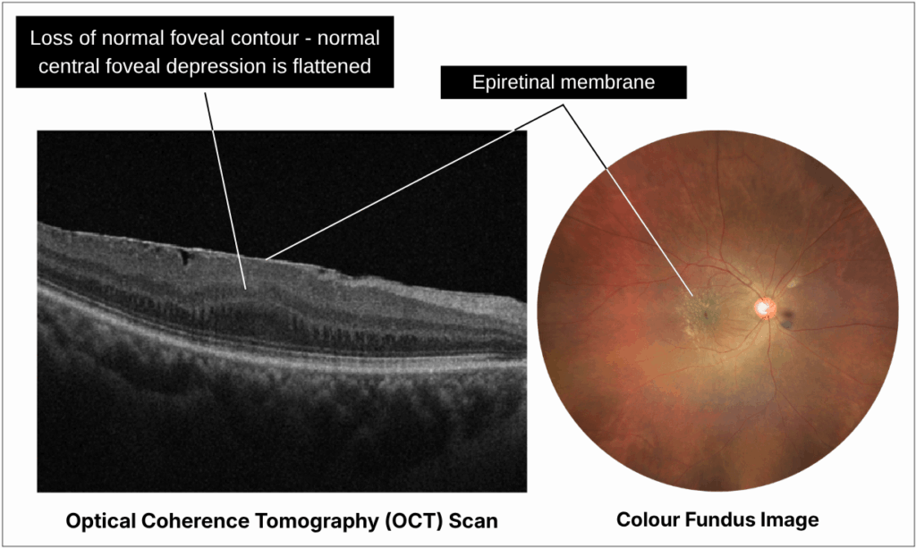
An epiretinal membrane is a thin layer of tissue that develops on the surface of the retina, usually over the macula, which is responsible for sharp central vision.
As the membrane thickens and contracts, it can pull on the macula and cause its normally smooth surface to become wrinkled or distorted. This can affect the way fine details, straight lines and shapes are seen. An epiretinal membrane is also known as a macular pucker.
Most epiretinal membranes develop as part of the natural ageing process. With age, the vitreous gel inside the eye gradually shrinks and separates from the retina, a process known as posterior vitreous detachment. During this process, cells can settle on the retinal surface and form a thin membrane over the macula.
In many cases, there is no other underlying eye condition responsible for the membrane.

Increasing age is the main risk factor for developing an epiretinal membrane. However, the condition may also be associated with:
An epiretinal membrane can also develop in an otherwise healthy eye with no previous eye condition or surgery.


Symptoms usually develop gradually and can vary considerably between individuals. Some epiretinal membranes remain stable with little effect on vision, while others may progressively interfere with everyday activities.
As the membrane tightens over the macula, vision may become blurred or distorted, with straight lines appearing wavy and differences in image size or clarity between the eyes.
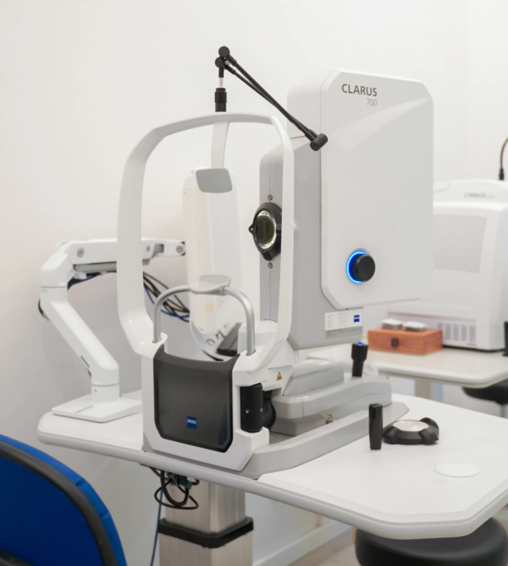
We use a combination of detailed eye examination and advanced retinal imaging to assess the macula, retina, optic nerve, and retinal blood vessels. These tests allow us to identify structural and functional changes, determine the type and severity of the condition, and establish a clear baseline for ongoing monitoring and treatment planning.
Measures how clearly you can see and helps identify any reduction in central vision.
Captures detailed images of the retina and macula to document the appearance and extent of the epiretinal membrane and monitor for changes over time.
Provides detailed cross-sectional images of the macula to identify an epiretinal membrane and assess the degree of retinal wrinkling, thickening or distortion it is causing.
Not every epiretinal membrane requires treatment. If the membrane is mild and has little effect on vision, it may simply be monitored with regular eye examinations and retinal scans. Eye drops, medications and nutritional supplements cannot remove the membrane or reverse the pulling on the retina.
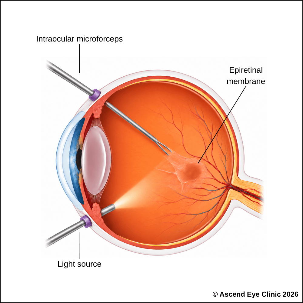
When blurred or distorted vision begins to interfere with daily activities, surgery is performed to remove the epiretinal membrane and release the traction on the macula. This allows the macula to gradually return towards a more normal shape, helping reduce distortion and improve central vision.
Vitrectomy is usually performed as an outpatient procedure, with no overnight hospital stay required. Fine microsurgical instruments are introduced through tiny openings in the eye to remove the vitreous gel and access the macula. The epiretinal membrane is then carefully peeled away, together with a very thin natural layer on the surface of the retina called the internal limiting membrane. Removing this layer helps ensure that residual membrane cells are cleared and reduces the likelihood of another epiretinal membrane developing in the same area.
The eye is usually left filled with fluid, although an air or gas bubble may be placed depending on the findings during surgery. If used, the bubble gradually dissolves and is naturally replaced by fluid within the eye.
Vision improves gradually as the eye heals. The final result depends on how significantly and how long the macula has been affected, and vision may not return completely to normal.
After surgery, we will monitor the eye closely with follow-up examinations and OCT scans to assess how the macula is healing and track changes in vision. Recovery is gradual, and the retina continues to settle over the weeks and months following surgery.
From your initial consultation to treatment and follow-up care, our team will guide you through every stage of your journey. We will take the time to explain your condition, answer your questions and help you feel informed and supported throughout your care.

Your consultation begins with a detailed assessment of your vision and eye health. Depending on your condition, this may include measurements of your vision and eye pressure, examination of the front and back of the eye, and advanced diagnostic imaging.
Our team will guide you through each test and explain what is being assessed. Your results will then be carefully reviewed to understand your individual needs and determine whether monitoring or treatment may be recommended.
Every eye and every patient is different. Your treatment plan will be tailored according to your examination findings, visual needs, general health and lifestyle.
We will explain the recommended treatment, its expected benefits, possible risks and any suitable alternatives in a clear and straightforward manner. You will have time to ask questions and consider your options before deciding how you would like to proceed.
Before your treatment, our team will guide you through the necessary preparations, paperwork and medication instructions. Where applicable, we will explain how to use your prescribed eye drops and how to care for your eyes afterwards.
We understand that undergoing a procedure can feel unfamiliar or worrying. Our team will be available to answer your questions and ensure that you know what to expect before the day of your treatment.
On the day of your procedure, you will arrive at the hospital or surgical centre where your operation has been scheduled. The surgical team will help you prepare and explain what will happen at each stage.
Anaesthetic eye drops or other forms of anaesthesia may be used, depending on the procedure, to keep you comfortable. Your doctor and the surgical team will support and reassure you throughout the operation.
Most eye surgeries are performed as day procedures, although the duration and recovery process will depend on the treatment you receive. Your vision may be temporarily blurred afterwards, so you should arrange for someone to accompany you home and should not drive unless your doctor has advised that it is safe to do so.
We will provide clear instructions on how to care for your eyes, use any prescribed medication and manage your activities during recovery. We will also explain which symptoms are expected and when you should contact the clinic for further advice.
Follow-up appointments allow us to monitor your vision, assess your recovery and identify any concerns at an early stage. Our team remains accessible should you have any questions or concerns throughout your recovery.
If straight lines appear wavy, objects look distorted or your central vision has become less clear, a detailed retinal assessment can help identify the cause.
Book a consultation at Ascend Eye Clinic for a comprehensive eye examination and advanced retinal imaging. We will explain your findings clearly and discuss whether monitoring or treatment may be appropriate.
An epiretinal membrane contracts on the surface of the macula and distorts its normal structure. Because the macula is responsible for detailed central vision, this distortion can make straight lines appear bent or wavy and cause letters or objects to look irregular.
Not necessarily. Many epiretinal membranes remain stable for years, while others gradually become thicker and create increasing traction on the macula. We monitor the membrane and the shape of the macula with OCT scans to identify meaningful progression.
Yes. An epiretinal membrane can develop in both eyes, although one eye is often more significantly affected than the other. When assessing one eye, we routinely examine the other eye for early changes as well.
Common symptoms include blurred central vision, wavy or distorted lines, difficulty reading fine print and reduced clarity when looking at faces or detailed objects. Some patients also notice that images appear slightly different in size or shape between the two eyes.
No. An epiretinal membrane is a layer of tissue growing over the surface of the macula, whereas a macular hole is an actual opening in the central retina. Both affect the macula and can cause distorted or reduced central vision, but they are different conditions with different structural changes on OCT.
No. An epiretinal membrane does not cause eye pain because it affects the retina at the back of the eye. The main changes are visual, particularly blurring and distortion of central vision.
Yes. Most epiretinal membranes are associated with normal age-related changes in the vitreous gel inside the eye. As the vitreous separates from the retina with age, cells can collect on the retinal surface and develop into a thin membrane over the macula.
Yes, particularly when significant traction has distorted the macula for a prolonged period. An epiretinal membrane generally affects central detail rather than causing complete blindness. Treating a visually significant membrane can relieve the traction and improve vision, although longstanding retinal changes can limit the final recovery.
We assess both how the membrane is affecting your vision and how much it has altered the macula. Visual acuity testing measures central vision, while OCT provides a detailed cross-sectional scan of the macula and shows the membrane, retinal wrinkling, thickening and traction.
No. A mild epiretinal membrane that is not significantly affecting vision can be monitored. Surgery is considered when distortion or reduced vision becomes significant enough to interfere with reading, driving, work or other everyday activities.
A mild, stable epiretinal membrane is commonly reviewed every 6 to 12 months with vision testing and OCT imaging. More regular reviews are needed when there is significant progression on the scan or a noticeable change in vision.
Surgery becomes appropriate when the epiretinal membrane is causing meaningful visual disturbance, particularly increasing distortion, reduced central clarity or difficulty with normal daily activities. We consider your symptoms together with your vision and OCT findings when recommending treatment.
Yes. When significant traction remains on the macula for a long period, the retinal structure can become increasingly distorted and the potential for complete visual recovery becomes lower. This is why we monitor progression carefully and recommend surgery at an appropriate stage rather than waiting until vision has deteriorated substantially.
The aim of surgery is to improve central vision and reduce distortion, but vision does not always return completely to normal. The amount of recovery is influenced by how much the macula was distorted before surgery and how long those changes had been present.
Visual recovery is gradual. Improvement usually becomes increasingly noticeable over the first few months and can continue for 6 to 12 months as the macula slowly remodels after the traction has been released.
Not routinely. In many epiretinal membrane operations, the eye can be left filled with fluid after the membrane has been removed. An air or gas bubble is used when there is a specific reason for additional internal support during surgery.
No. Epiretinal membrane surgery is generally performed as day surgery. We will monitor you after the operation, and once you are comfortable and medically stable, you can return home on the same day.
Recurrence is uncommon after successful membrane peeling. During surgery, we can also remove a very thin layer from the surface of the retina to further reduce the chance of another membrane forming in the same area.
Vitrectomy accelerates cataract development in eyes that still have their natural lens, particularly in older adults. If a cataract is already present, we will assess its significance and discuss whether cataract surgery should be planned separately or together with the retinal procedure.
Light everyday activities can usually resume relatively soon after surgery, while strenuous exercise and heavy lifting require a longer period of recovery. Driving should only resume once vision has recovered sufficiently and meets the required driving standard. We will review the eye after surgery and guide you on when each activity can be safely restarted.
Hi, I would like to book a consultation with Dr Errol Chan from Ascend Eye Clinic
