
Central serous chorioretinopathy (CSCR) causes fluid to collect beneath the retina, disrupting the macula and affecting the quality of central vision. Detailed retinal imaging helps us assess the extent of fluid, monitor recovery, and guide the most suitable treatment strategy.
Central serous chorioretinopathy, or CSCR, is a retinal condition in which fluid collects beneath the retina, usually at the macula. This causes a small area of the retina to lift away from the tissue beneath it, which may blur or distort central vision.
CSCR occurs when the blood vessels beneath the retina become abnormally permeable, causing fluid to leak through the retinal pigment epithelium and collect beneath the retina. This fluid accumulation can lift the central retina and affect vision.
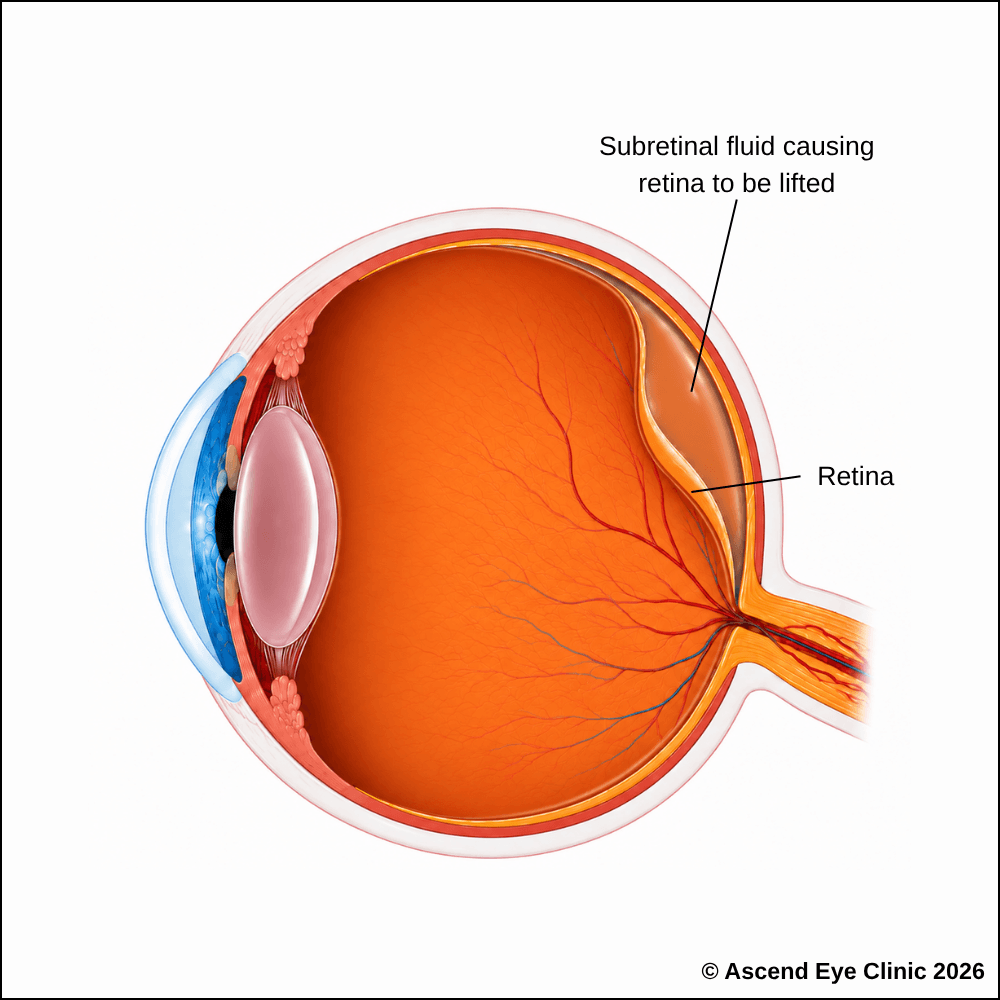

CSCR can affect adults of any age but is most frequently seen in young and middle-aged adults. It is also diagnosed more commonly in men than women.
CSCR has been associated with several factors, including:


Symptoms of CSCR often include blurred or reduced central vision, distortion of straight lines, reduced contrast or colour intensity, and objects appearing smaller or further away in the affected eye.
Symptoms vary depending on the amount and location of fluid beneath the retina. Many first episodes improve as the fluid resolves, while persistent or recurrent fluid can lead to lasting central vision changes.
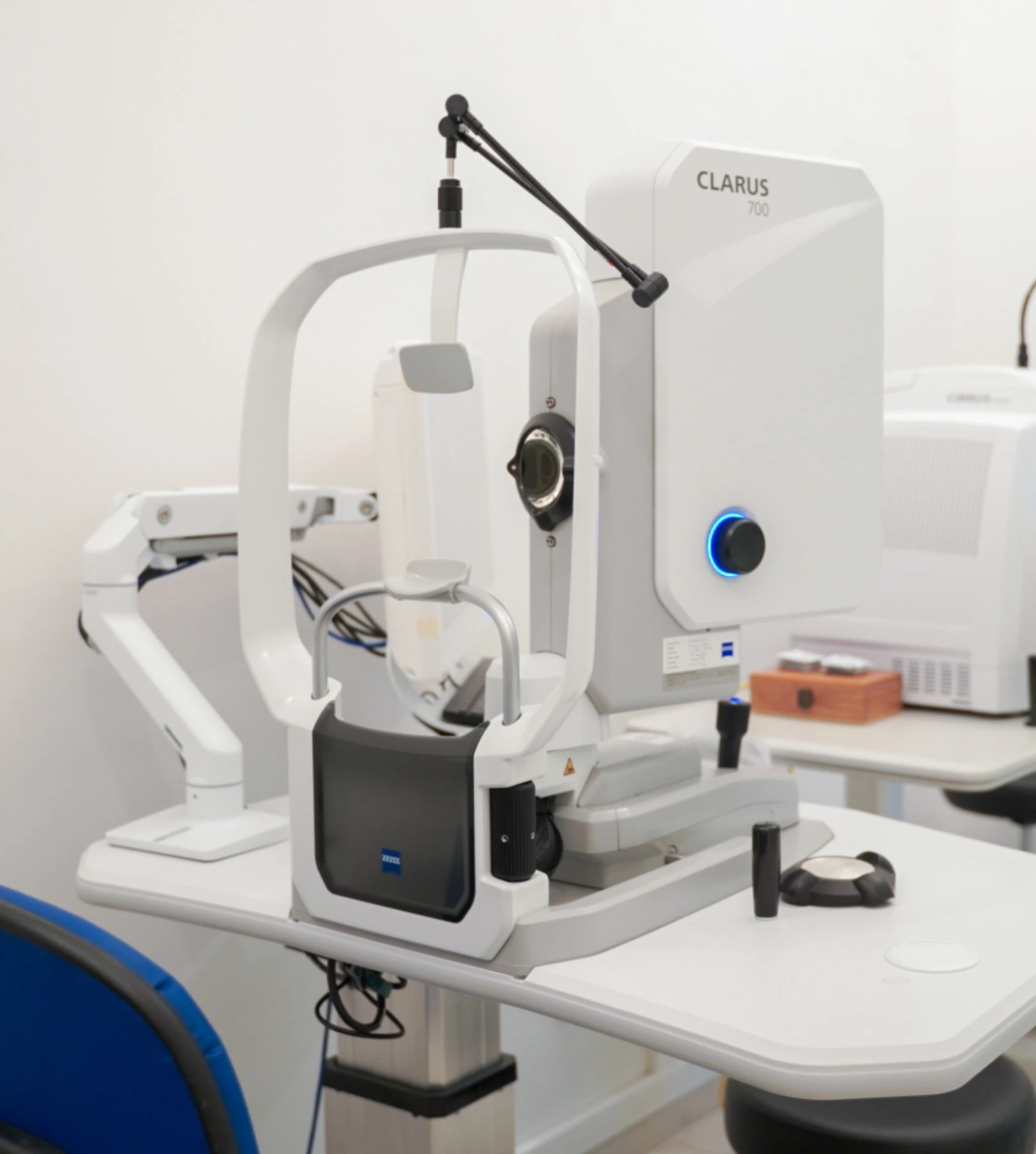
We use a combination of detailed eye examination and advanced retinal imaging to assess the macula, retina, optic nerve, and retinal blood vessels. These tests allow us to identify structural and functional changes, determine the type and severity of the condition, and establish a clear baseline for ongoing monitoring and treatment planning.
Measures how clearly you can see and helps identify any reduction in central vision.
Captures detailed images of the retina to identify changes associated with central serous chorioretinopathy, including areas of retinal pigment epithelium disturbance and other retinal abnormalities.
This is useful in chronic CSCR as this will highlight areas of the retina which have been exposed to longstanding fluid.
Uses fluorescein dye and a series of retinal photographs to help identify the source of the leak.
Provides cross-sectional images of the retina to assess the macula and individual retinal layers for swelling, fluid or other structural changes.
Treatment may be considered earlier if the fluid persists, vision is significantly affected, the condition repeatedly returns or clear central vision is important for the patient’s occupation.
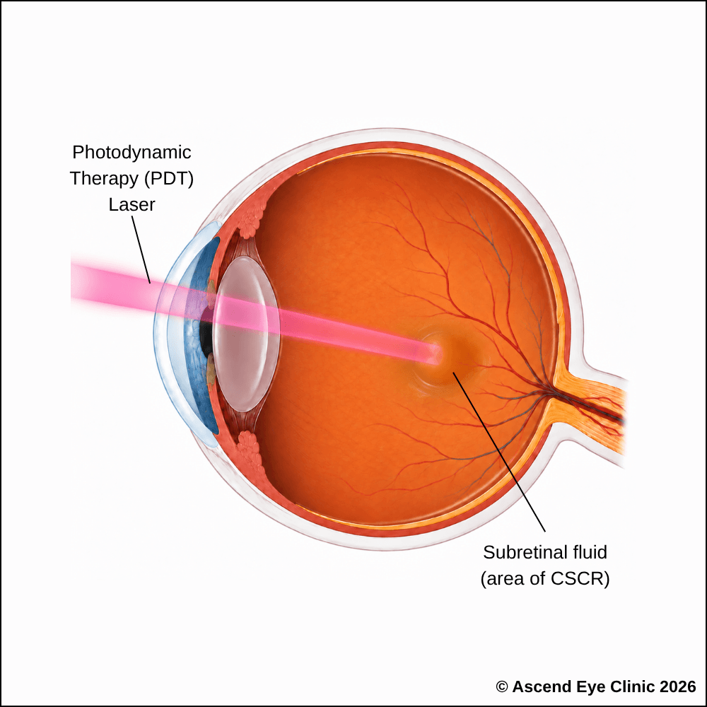
Photodynamic therapy (PDT) is a targeted treatment used for persistent or recurrent CSCR, particularly when ongoing leakage from the choroid causes subretinal fluid to remain beneath the retina.
PDT is performed as an outpatient procedure and does not require a hospital stay. Treatment begins with an intravenous injection of verteporfin, a light-sensitive medication that circulates through the bloodstream and reaches the choroidal blood vessels beneath the retina. A specialised low-energy laser is then directed precisely at the area of abnormal choroidal leakage. The laser activates the verteporfin, producing a local reaction that reduces excessive permeability of the choroidal vessels while limiting damage to the surrounding retina.
By reducing this abnormal leakage, PDT allows the accumulated subretinal fluid to gradually resolve and the retina to settle back into its normal position. This helps restore the normal structure of the macula, stabilise vision and reduce the likelihood of persistent or recurrent fluid.
From your initial consultation to treatment and follow-up care, our team will guide you through every stage of your journey. We will take the time to explain your condition, answer your questions and help you feel informed and supported throughout your care.

Your consultation begins with a detailed assessment of your vision and eye health. Depending on your condition, this may include measurements of your vision and eye pressure, examination of the front and back of the eye, and advanced diagnostic imaging.
Our team will guide you through each test and explain what is being assessed. Your results will then be carefully reviewed to understand your individual needs and determine whether monitoring or treatment may be recommended.
Every eye and every patient is different. Your treatment plan will be tailored according to your examination findings, visual needs, general health and lifestyle.
We will explain the recommended treatment, its expected benefits, possible risks and any suitable alternatives in a clear and straightforward manner. You will have time to ask questions and consider your options before deciding how you would like to proceed.
Before your treatment, our team will guide you through the necessary preparations, paperwork and medication instructions. Where applicable, we will explain how to use your prescribed eye drops and how to care for your eyes afterwards.
We understand that undergoing a procedure can feel unfamiliar or worrying. Our team will be available to answer your questions and ensure that you know what to expect before the day of your treatment.
On the day of your procedure, you will arrive at the hospital or surgical centre where your operation has been scheduled. The surgical team will help you prepare and explain what will happen at each stage.
Anaesthetic eye drops or other forms of anaesthesia may be used, depending on the procedure, to keep you comfortable. Your doctor and the surgical team will support and reassure you throughout the operation.
Most eye surgeries are performed as day procedures, although the duration and recovery process will depend on the treatment you receive. Your vision may be temporarily blurred afterwards, so you should arrange for someone to accompany you home and should not drive unless your doctor has advised that it is safe to do so.
We will provide clear instructions on how to care for your eyes, use any prescribed medication and manage your activities during recovery. We will also explain which symptoms are expected and when you should contact the clinic for further advice.
Follow-up appointments allow us to monitor your vision, assess your recovery and identify any concerns at an early stage. Our team remains accessible should you have any questions or concerns throughout your recovery.
If you have noticed blurred, distorted or dim central vision, a detailed retinal assessment can help identify the cause.
Book a consultation at Ascend Eye Clinic for a comprehensive examination and advanced retinal imaging. We will explain your findings clearly and advise whether monitoring or treatment is recommended.
No. Many first episodes improve naturally over several weeks or months, but fluid may persist or repeatedly return in some patients. Treatment may be recommended when recovery is delayed or there is a risk of lasting retinal damage.
This is assessed based on how long the fluid has been present and the appearance of the retina on OCT and other imaging. Chronic CSCR may show persistent fluid, widespread changes beneath the retina or signs of damage from previous episodes.
Many patients regain good visual acuity after the fluid resolves. However, subtle distortion, reduced contrast or changes in colour perception may remain, particularly when fluid has persisted or CSCR has occurred more than once.
Yes. CSCR can recur in the same eye and may later affect the other eye. Further assessment is recommended if blurred, dim or distorted central vision returns.
No, you should not stop the steroid medication on your own accord as the steroids may have been given for other medical purposes. Steroids can trigger or worsen CSCR, but the prescribed steroid medication should not be stopped suddenly. It is best to review it with the doctor who prescribed it so that it can be safely reduced, changed or discontinued where appropriate. Steroids may be present in tablets, injections, inhalers, nasal sprays and skin creams.
Reducing stress may be beneficial for general health and may help address a possible trigger, but it does not guarantee that the retinal fluid will resolve. Regular retinal imaging remains important even when stress levels improve.
CSCR causes a localised pocket of fluid beneath the sensory retina, usually around the macula. It is different from the more extensive retinal detachment caused by a retinal tear, although both involve separation of retinal layers.
Symptoms may improve before all the fluid has disappeared. OCT allows the ophthalmologist to detect small amounts of remaining fluid and determine whether the retina is continuing to recover.
No. PDT is an outpatient laser treatment and no instruments are placed inside the eye. A light-sensitive medication is administered through a vein before a controlled light treatment is applied to the affected area.
Most patients do not experience pain from the laser treatment. You may feel mild discomfort when the intravenous line is placed, and your eyes may be temporarily sensitive to light following pupil dilation.
The medication circulates to the blood vessels beneath the retina and becomes active only when exposed to a specific laser light. This allows the ophthalmologist to target the area responsible for abnormal leakage.
The medication temporarily increases sensitivity to light. Direct sunlight and strong indoor lighting can cause a photosensitivity reaction, so you will be given instructions on protective clothing, sunglasses and how long additional precautions are required.
The fluid does not usually disappear immediately. It may reduce gradually over the following weeks, and OCT scans will be used to assess how the retina is responding.
PDT aims to reduce leakage and help the fluid resolve, but the amount of visual improvement varies. Vision may remain limited if the macula has already developed lasting changes from prolonged or recurrent CSCR.
Yes, further PDT may be considered if fluid persists or returns. The decision will depend on previous treatment, retinal imaging and the condition of the surrounding tissue. Reduced-setting PDT has demonstrated better anatomical outcomes than micropulse laser in chronic CSCR, although treatment must still be tailored to each eye.
Hi, I would like to book a consultation with Dr Errol Chan from Ascend Eye Clinic
