
The retina plays a vital role in vision, and retinal conditions can cause blurred or distorted vision, flashes, floaters, or dark areas in the visual field.
At Ascend Eye Clinic, we provide comprehensive retinal assessment using detailed eye examinations and high-resolution imaging to establish an accurate diagnosis and guide appropriate monitoring, medical, laser or surgical treatment.
Retinal conditions are a group of eye disorders that affect the retina, the light-sensitive layer at the back of the eye responsible for capturing visual information. Some retinal conditions develop gradually and may not cause noticeable symptoms initially, while others can cause sudden changes in vision, requiring urgent treatment.
Conditions commonly assessed and treated include:
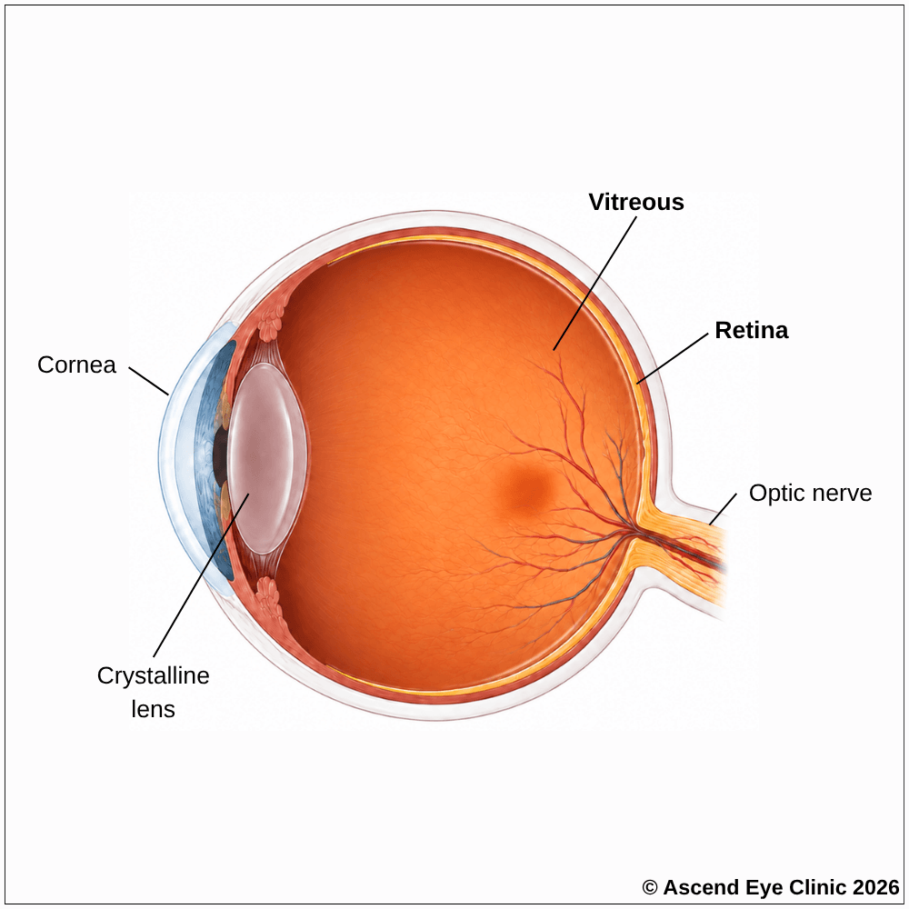
Retinal conditions can affect central vision, peripheral vision, or both. Symptoms either develop gradually or appear suddenly depending on the underlying conditions.

Associated with posterior retinal detachment, retinal tears or retinal detachment

Commonly seen in macular degeneration, epiretinal membrane, or macular swelling

A warning sign of retinal detachment
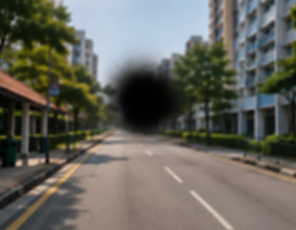
May occur with retinal vein occlusion, vitreous haemorrhage or other retinal vascular conditions

May be associated with inherited retinal conditions such as retinitis pigmentosa
A retinal assessment combines examination with detailed imaging to evaluate both the structure and function of the eye.
Measures how clearly you can see and helps identify any reduction in central vision.
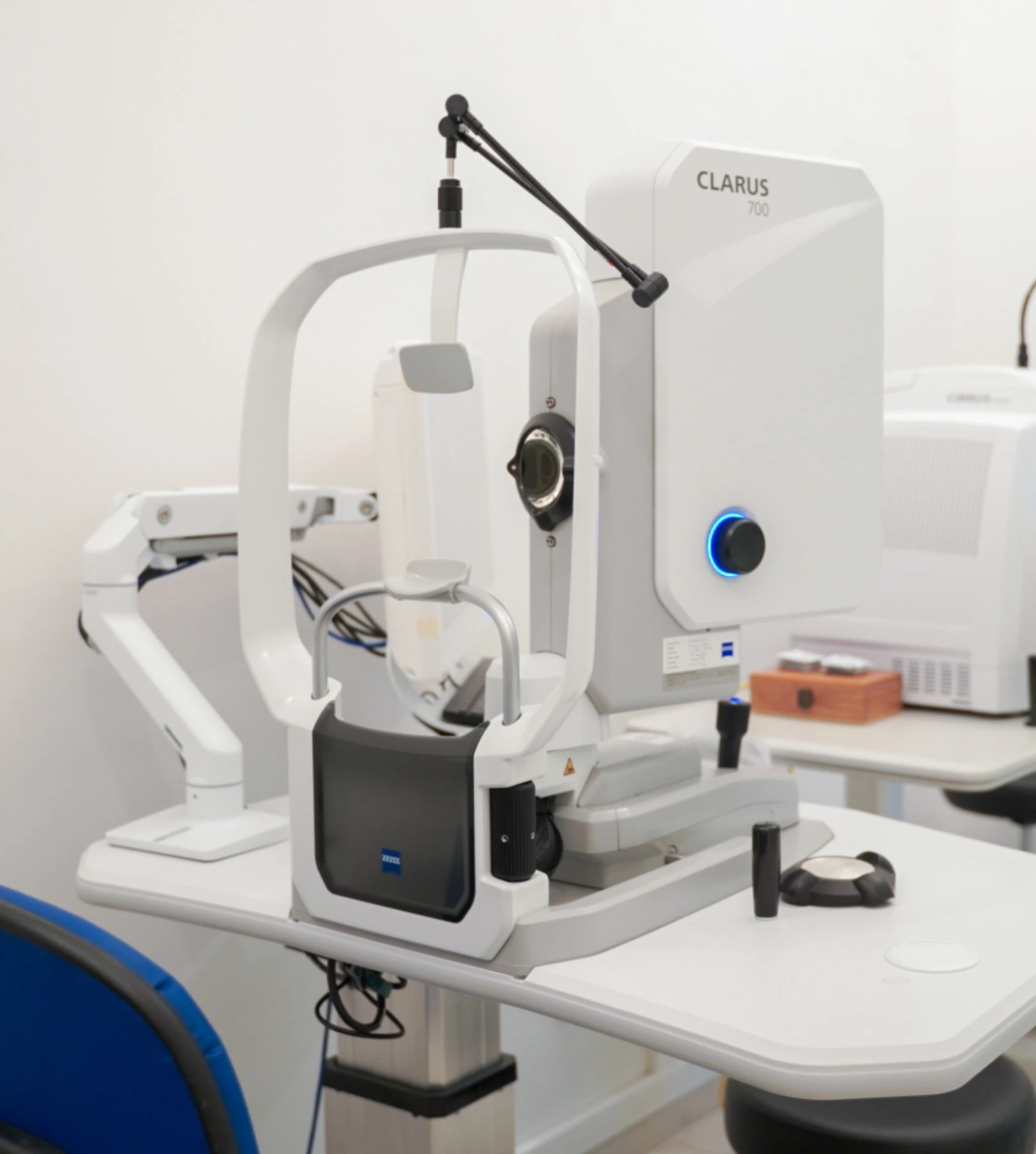
Captures detailed images of the retina and retinal blood vessels to identify abnormalities and signs of retinal diseases.
Uses fluorescein dry and a series of retinal photographs to assess blood flow through the retinal circulation to identify leakage, blocked blood vessels, and abnormal new blood vessels.
Uses indocyanine green dry to provide detailed imaging of the deeper blood vessels beneath the retina, particularly the choroidal circulation, helping identify abnormalities that may not be clearly visible with fundus fluorescein angiography.
Provides cross-sectional images of the retina to assess the macula and individual retinal layers for swelling, fluid or other structural changes.
Provides a non-invasive imaging of blood flow within the retinal and choroidal vessels at the macula, without the use of injected dye to identify areas of reduced circulation and abnormal blood vessel growth.
Maps your central and peripheral visual field to identify areas of reduced or missing vision that can occur with certain retinal or optic nerve conditions.
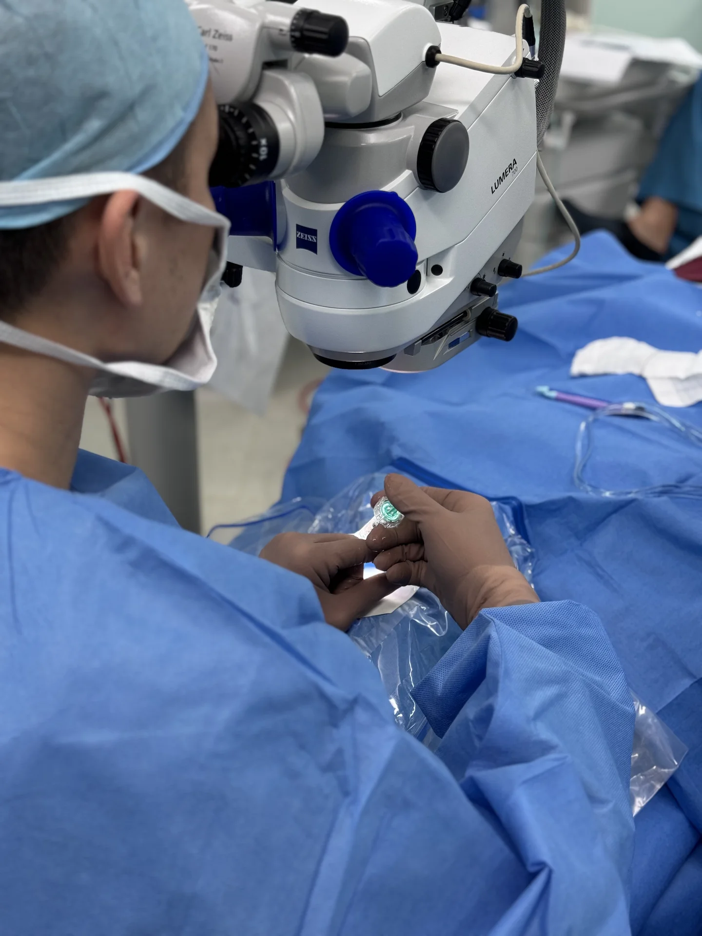
Treatment is selected according to the diagnosis, severity of the condition, and its effect on vision. Some stable or early retinal changes may only require regular monitoring, while others require prompt treatment to prevent further deterioration
At Ascend Eye Clinic, your findings and treatment options will be explained clearly. Where treatment is recommended, the approach will be tailored to your condition, visual needs and overall eye health.
From your initial consultation to treatment and follow-up care, our team will guide you through every stage of your journey. We will take the time to explain your condition, answer your questions and help you feel informed and supported throughout your care.

Your consultation begins with a detailed assessment of your vision and eye health. Depending on your condition, this may include measurements of your vision and eye pressure, examination of the front and back of the eye, and advanced diagnostic imaging.
Our team will guide you through each test and explain what is being assessed. Your results will then be carefully reviewed to understand your individual needs and determine whether monitoring or treatment may be recommended.
Every eye and every patient is different. Your treatment plan will be tailored according to your examination findings, visual needs, general health and lifestyle.
We will explain the recommended treatment, its expected benefits, possible risks and any suitable alternatives in a clear and straightforward manner. You will have time to ask questions and consider your options before deciding how you would like to proceed.
Before your treatment, our team will guide you through the necessary preparations, paperwork and medication instructions. Where applicable, we will explain how to use your prescribed eye drops and how to care for your eyes afterwards.
We understand that undergoing a procedure can feel unfamiliar or worrying. Our team will be available to answer your questions and ensure that you know what to expect before the day of your treatment.
On the day of your procedure, you will arrive at the hospital or surgical centre where your operation has been scheduled. The surgical team will help you prepare and explain what will happen at each stage.
Anaesthetic eye drops or other forms of anaesthesia may be used, depending on the procedure, to keep you comfortable. Your doctor and the surgical team will support and reassure you throughout the operation.
Most eye surgeries are performed as day procedures, although the duration and recovery process will depend on the treatment you receive. Your vision may be temporarily blurred afterwards, so you should arrange for someone to accompany you home and should not drive unless your doctor has advised that it is safe to do so.
We will provide clear instructions on how to care for your eyes, use any prescribed medication and manage your activities during recovery. We will also explain which symptoms are expected and when you should contact the clinic for further advice.
Follow-up appointments allow us to monitor your vision, assess your recovery and identify any concerns at an early stage. Our team remains accessible should you have any questions or concerns throughout your recovery.
Symptoms such as new floaters, flashes of light, distortion, blurred vision or a dark area in your vision may be related to a retinal condition. Some retinal problems can progress quickly, even when symptoms initially seem mild.
Book a consultation at Ascend Eye Clinic for a detailed retinal assessment. We will thoroughly examine your eyes, perform the necessary imaging and explain whether monitoring, treatment or surgery is recommended.
Retinal conditions can cause blurred or distorted vision, flashes, new floaters, missing areas of vision, reduced peripheral vision or a dark curtain or shadow across part of the visual field. Some retinal conditions cause no symptoms in their early stages, which is why retinal examination and imaging are important when relevant risk factors are present.
No. Floaters are common and often arise from normal age-related changes in the vitreous gel inside the eye. However, a sudden increase in floaters, particularly when accompanied by flashes or a shadow in the vision, requires prompt assessment to exclude a retinal tear or retinal detachment.
Seek prompt assessment if flashes or floaters appear suddenly, increase significantly, or are accompanied by blurred vision, a dark shadow or loss of part of your visual field. These symptoms can occur when the vitreous pulls on the retina and can indicate a retinal tear.
A dark curtain, veil or shadow spreading across part of the vision is an important warning symptom of retinal detachment. This requires urgent assessment because a detached retina can lead to permanent loss of vision if it progresses.
Wavy or distorted lines can indicate a problem affecting the macula, the central part of the retina responsible for detailed vision. Conditions such as an epiretinal membrane, macular degeneration, macular swelling or a macular hole can produce this type of visual distortion.
Sudden loss or blurring of vision can result from several retinal problems, including retinal detachment, retinal vascular occlusion, bleeding into the vitreous or sudden changes affecting the macula. A rapid change in vision should be assessed promptly so the cause can be identified.
Yes. Early diabetic retinopathy, retinal vascular changes and some peripheral retinal abnormalities can develop before a person notices any change in vision. Detailed retinal examination and imaging allow these changes to be detected before they significantly affect sight.
Yes. Many retinal conditions can develop in one eye independently of the other. Even when only one eye is symptomatic, we assess both eyes because certain retinal diseases and risk factors can affect both sides.
Retinal problems can result from ageing, changes in the vitreous, diabetes, abnormal retinal blood vessels, high myopia, inflammation, trauma and inherited retinal disorders. The underlying cause varies considerably, so establishing an accurate diagnosis is important.
Important risk factors include increasing age, diabetes, high myopia, previous retinal problems, eye trauma, previous eye surgery and a family history of certain retinal diseases. Some systemic conditions that affect the blood vessels can also affect the retinal circulation.
Yes. The retina and vitreous undergo natural changes with age. These changes increase the likelihood of conditions such as posterior vitreous detachment, retinal tears, macular degeneration and epiretinal membrane, particularly from middle age onwards.
We begin with a detailed history and eye examination, including visual acuity and a dilated examination of the retina. Retinal imaging is then used to examine specific structures in greater detail and identify abnormalities that are not always apparent from symptoms alone.
For a comprehensive retinal assessment, we often dilate the pupils to obtain a wider and clearer view of the retina, including the peripheral retina. Vision can be temporarily blurred and more sensitive to light after dilation, particularly for near tasks.
Depending on the condition being investigated, we use tests such as optical coherence tomography (OCT), wide-field retinal photography and other specialised retinal imaging. These provide detailed views of the macula, retinal layers, optic nerve and peripheral retina, allowing subtle abnormalities to be documented and followed over time.
Yes. The retina contains highly specialised nerve tissue, and significant damage can result in permanent visual loss. Early diagnosis is therefore important, particularly when symptoms develop suddenly or when risk factors for retinal disease are present.
Hi, I would like to book a consultation with Dr Errol Chan from Ascend Eye Clinic
