
Uveitis can affect different structures in the eye and cause redness, pain, light sensitivity, floaters or blurred vision. Early treatment is important to control inflammation and reduce the risk of complications that may affect vision.
Uveitis is inflammation inside the eye. It can affect different structures within the eye and may involve one or both eyes. Some people experience a single episode, while others have inflammation that returns or persists over time.
Structures that can be affected include:
Uveitis is associated with infections, autoimmune or inflammatory conditions, eye injuries, and previous eye surgery. In many cases, however, no specific cause is identified.
Early diagnosis and treatment are important, as uncontrolled inflammation can lead to complications such as cataract, glaucoma, macular swelling, and retinal damage.
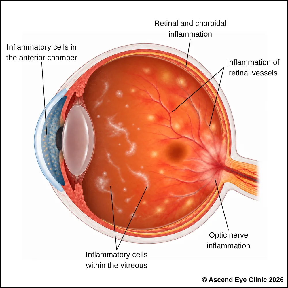
The symptoms vary according to the area of the eye affected. Anterior uveitis commonly causes a painful, red, and light-sensitive eye. Intermediate and posterior uveitis more often cause blurred vision and floaters, sometimes without obvious redness or pain.
Common symptoms include:

Diagnosing uveitis involves a careful examination of the eye to determine where the inflammation is occurring, how active it is and whether any complications are present. Additional investigations may be performed depending on the type of uveitis and the findings during your examination.
Measures how clearly you can see and helps identify any changes in vision caused by inflammation.
Allows detailed examination of the front of the eye to look for signs of inflammation involving structures such as the iris and ciliary body.
Provides a detailed view of the vitreous, retina, choroid and optic nerve to identify inflammation affecting the middle or back of the eye.
Uses fluorescein dye and a series of retinal photographs to identify areas of inflammation, leakage or changes in the retinal blood vessels.
Provides detailed imaging of the deeper choroidal circulation and may be useful when inflammation involving the choroid is suspected.
Provides detailed cross-sectional images of the retina and macula to detect swelling, fluid or other changes associated with inflammation.
Provides detailed images of the retinal and choroidal blood vessels without the use of dye, helping to identify areas of reduced blood flow or macular ischaemia associated with uveitis.
In selected cases, additional investigations may be recommended to look for an underlying infection, autoimmune or inflammatory condition associated with uveitis.
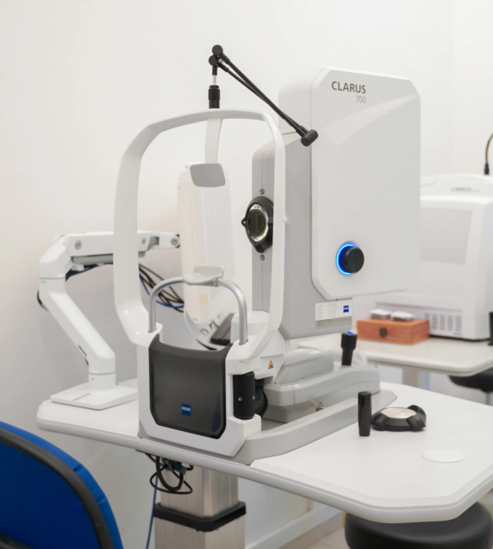
Not every patient requires every investigation. The tests recommended will depend on the location and severity of the inflammation, your symptoms, examination findings and medical history.
Treatment for uveitis focuses on controlling inflammation, relieving symptoms, protecting vision and treating any underlying cause or complication. The treatment approach depends on which part of the eye is affected, the severity of inflammation and whether the uveitis is associated with an infection, autoimmune condition or other disease.
Steroid eye drops are commonly used to reduce inflammation in anterior uveitis. Dilating eye drops are often prescribed alongside them to relax the muscles inside the eye, relieve discomfort and reduce the risk of the iris sticking to the lens.
For recurrent, persistent or more severe uveitis, immunosuppressive medication is used to control inflammation and reduce reliance on long-term steroid treatment. These medications work by modifying the immune response that is driving inflammation inside the eye.
Treatment is coordinated with the relevant medical specialist where systemic monitoring is required, with regular blood tests and follow-up to assess treatment response and medication safety.

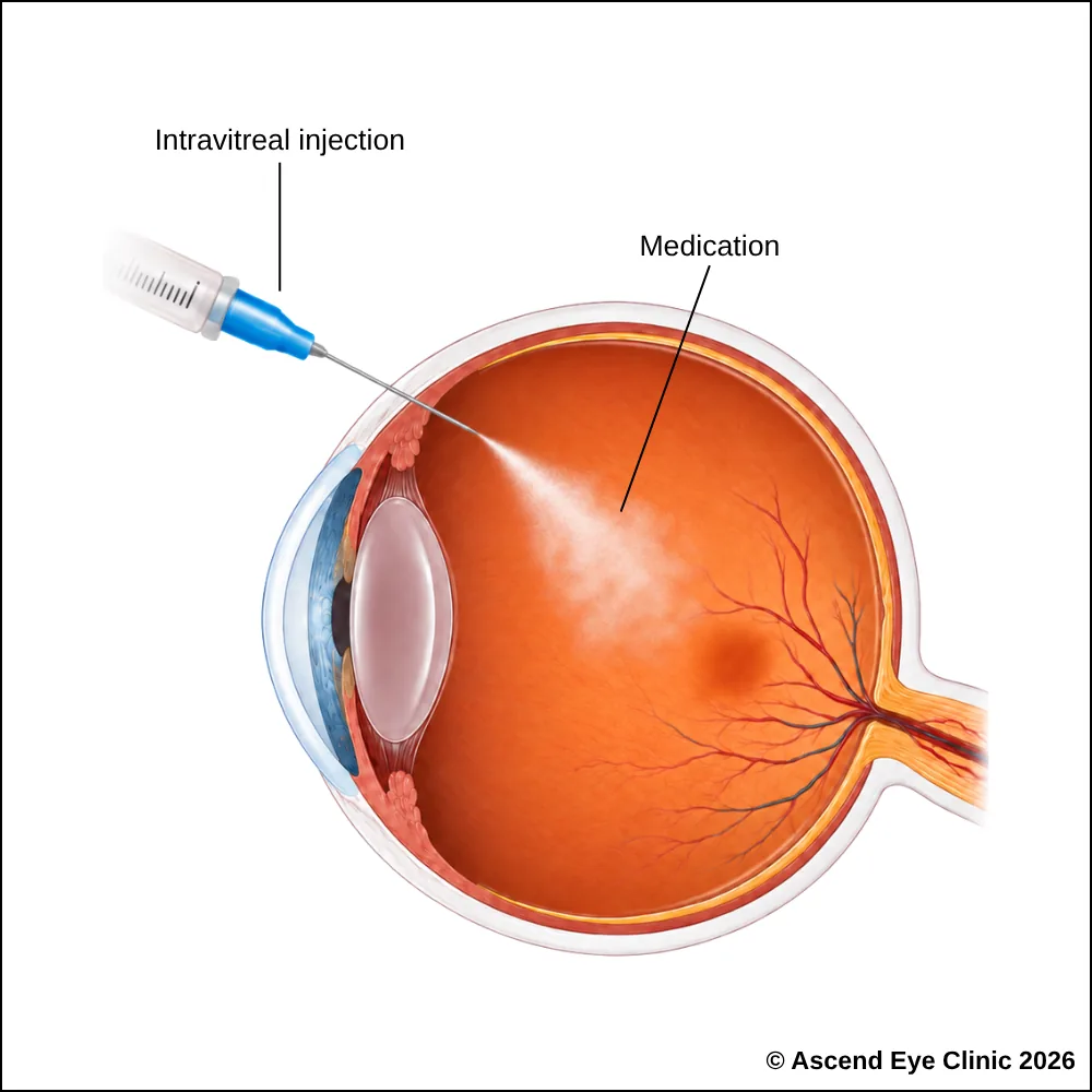
An intravitreal injection delivers medication directly into the vitreous to deliver treatment closer to the retina and other structures at the back of the eye.
Steroid injections or sustained-release steroid implants such as Ozurdex® are used to control inflammation and treat uveitic macular oedema. For infective uveitis, intravitreal antibiotics or antiviral medications are injected directly into the eye to treat the underlying infection. Anti-VEGF injections are used when uveitis is associated with complications such as abnormal retinal blood vessel leakage.
An intravitreal injection is performed as a quick outpatient procedure with careful preparation to keep the eye comfortable throughout. Anaesthetic drops are used to thoroughly numb the eye before the injection. Although the idea of an injection into the eye can sound daunting, the injection itself takes only a few seconds, much like a vaccination procedure, and most people feel only very mild discomfort.
Learn more about intravitreal injections here.
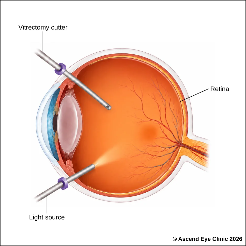
Vitrectomy removes the vitreous gel from inside the eye. It is used when persistent or severe vitritis causes significant visual disturbance, when complications involving the retina require surgical treatment, or when the cause of inflammation remains unclear.
Vitrectomy is a keyhole surgery performed in an operating theatre under sedation or general anaesthesia. This is a keyhole surgery, where fine instruments are introduced through tiny ports on the surface of the eye. A sample of the vitreous is collected and sent for laboratory analysis to investigate infections, inflammatory conditions and proliferative disorders such as intraocular lymphoma. This helps establish the diagnosis and guide further treatment when the underlying cause of uveitis is uncertain.
Learn more about a vitrectomy here.
Repeated or prolonged inflammation inside the eye can lead to complications such as cataract and glaucoma.
Cataract develops when ongoing inflammation, together with prolonged steroid treatment in some patients, causes the natural lens in the eye to become progressively cloudy. Cataract surgery is performed when this clouding significantly affects vision and the inflammation has been brought under appropriate control. Learn more about cataract surgery here.
Uveitic glaucoma develops when inflammation interferes with the eye’s normal drainage system. Inflammatory cells and debris can obstruct the trabecular meshwork, while swelling, scarring or adhesions within the front of the eye can further reduce aqueous drainage. Steroid treatment can also increase eye pressure in susceptible patients. When raised eye pressure is not adequately controlled with medication or laser treatment, glaucoma surgery such as trabeculectomy or glaucoma drainage implant surgery is considered. Learn more about glaucoma treatment options here.
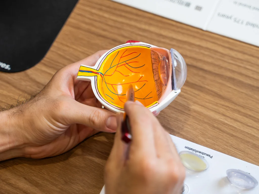
From your initial consultation to treatment and follow-up care, our team will guide you through every stage of your journey. We will take the time to explain your condition, answer your questions and help you feel informed and supported throughout your care.

Your consultation begins with a detailed assessment of your vision and eye health. Depending on your condition, this may include measurements of your vision and eye pressure, examination of the front and back of the eye, and advanced diagnostic imaging.
Our team will guide you through each test and explain what is being assessed. Your results will then be carefully reviewed to understand your individual needs and determine whether monitoring or treatment may be recommended.
Every eye and every patient is different. Your treatment plan will be tailored according to your examination findings, visual needs, general health and lifestyle.
We will explain the recommended treatment, its expected benefits, possible risks and any suitable alternatives in a clear and straightforward manner. You will have time to ask questions and consider your options before deciding how you would like to proceed.
Before your treatment, our team will guide you through the necessary preparations, paperwork and medication instructions. Where applicable, we will explain how to use your prescribed eye drops and how to care for your eyes afterwards.
We understand that undergoing a procedure can feel unfamiliar or worrying. Our team will be available to answer your questions and ensure that you know what to expect before the day of your treatment.
On the day of your procedure, you will arrive at the hospital or surgical centre where your operation has been scheduled. The surgical team will help you prepare and explain what will happen at each stage.
Anaesthetic eye drops or other forms of anaesthesia may be used, depending on the procedure, to keep you comfortable. Your doctor and the surgical team will support and reassure you throughout the operation.
Most eye surgeries are performed as day procedures, although the duration and recovery process will depend on the treatment you receive. Your vision may be temporarily blurred afterwards, so you should arrange for someone to accompany you home and should not drive unless your doctor has advised that it is safe to do so.
We will provide clear instructions on how to care for your eyes, use any prescribed medication and manage your activities during recovery. We will also explain which symptoms are expected and when you should contact the clinic for further advice.
Follow-up appointments allow us to monitor your vision, assess your recovery and identify any concerns at an early stage. Our team remains accessible should you have any questions or concerns throughout your recovery.
Uveitis requires prompt assessment to control inflammation and protect your vision. Book a consultation at Ascend Eye Clinic for a detailed eye examination and personalised treatment.
Uveitis is inflammation inside the eye. It can affect different parts of the eye and range from a single episode to a condition that returns or requires longer-term treatment.
Uveitis has many possible causes. It can be linked to autoimmune or inflammatory conditions, infections, previous eye injury or other medical conditions. In some patients, no definite underlying cause is found.
Uveitis needs proper assessment because persistent inflammation can damage structures inside the eye and affect vision. Early treatment helps control inflammation and reduce the risk of complications.
Yes. Uveitis can affect one eye or both eyes, either at the same time or at different times. The pattern often depends on the underlying cause.
Yes. Some people experience a single episode, while others have recurrent or chronic inflammation. Regular follow-up helps us detect a recurrence early and restart or adjust treatment promptly.
Yes. Uveitis can be associated with inflammatory or autoimmune conditions affecting other parts of the body, including the joints, skin or digestive system. We will ask about your general health and arrange further investigations when needed.
Not every patient requires extensive testing. Investigations are arranged when the pattern of inflammation, medical history or examination findings suggest an underlying cause that needs to be identified.
Treatment focuses on controlling inflammation and treating the underlying cause when one is identified. This commonly includes anti-inflammatory eye drops, pupil-dilating drops and, for more extensive inflammation, tablets, injections or other medications.
The recovery period depends on the type and severity of inflammation. Some episodes improve over several weeks, while others require longer treatment and closer monitoring before the eye becomes stable.
Many patients recover good vision once the inflammation settles. The final vision depends on how severe the inflammation was and whether complications such as retinal swelling, glaucoma, cataract or scarring developed.
Yes, severe or poorly controlled inflammation can lead to permanent visual damage. Prompt treatment and regular monitoring greatly reduce the risk by allowing us to control inflammation and manage complications early.
Inflammation inside the eye can remain active even after pain, redness or blurred vision have improved. Follow-up allows us to confirm that the inflammation has settled and to check the eye pressure, retina and other structures for complications.
No. Anti-inflammatory drops often need to be reduced gradually rather than stopped suddenly. Stopping treatment too early can allow the inflammation to return, so we will provide a clear schedule for reducing the medication.
Yes. The inflammation itself and some medications used to treat it can increase eye pressure. We check the pressure during follow-up and treat any significant rise to protect the optic nerve.
Seek prompt assessment if you develop a sudden drop in vision, increasing eye pain, marked redness, new flashes or floaters, severe light sensitivity or a significant worsening of your symptoms. These changes can indicate increasing inflammation or a complication that needs early treatment.
Hi, I would like to book a consultation with Dr Errol Chan from Ascend Eye Clinic
