
A choroidal naevus is a pigmented spot at the back of the eye that is often discovered during a routine eye examination. Detailed retinal imaging helps document its size, appearance and surrounding retina so that any changes can be identified over time and appropriate follow-up can be planned.
A choroidal naevus is a pigmented area in the choroid, the layer of tissue beneath the retina. It is made up of pigment-producing cells and is often described as being similar to a freckle or mole at the back of the eye. Most choroidal naevi are harmless, remain stable over time and do not affect vision, but regular imaging helps document their appearance and identify any changes that require closer assessment.
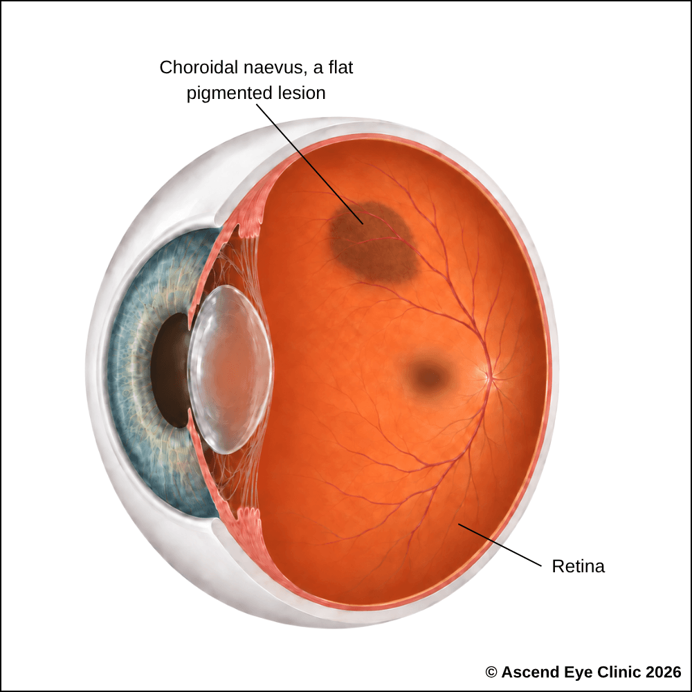
Most choroidal naevi do not cause symptoms. If new symptoms develop, such as blurred or distorted vision, flashes, floaters or reduced vision, this may indicate that the naevus has changed or is affecting the surrounding retina and should be reassessed.
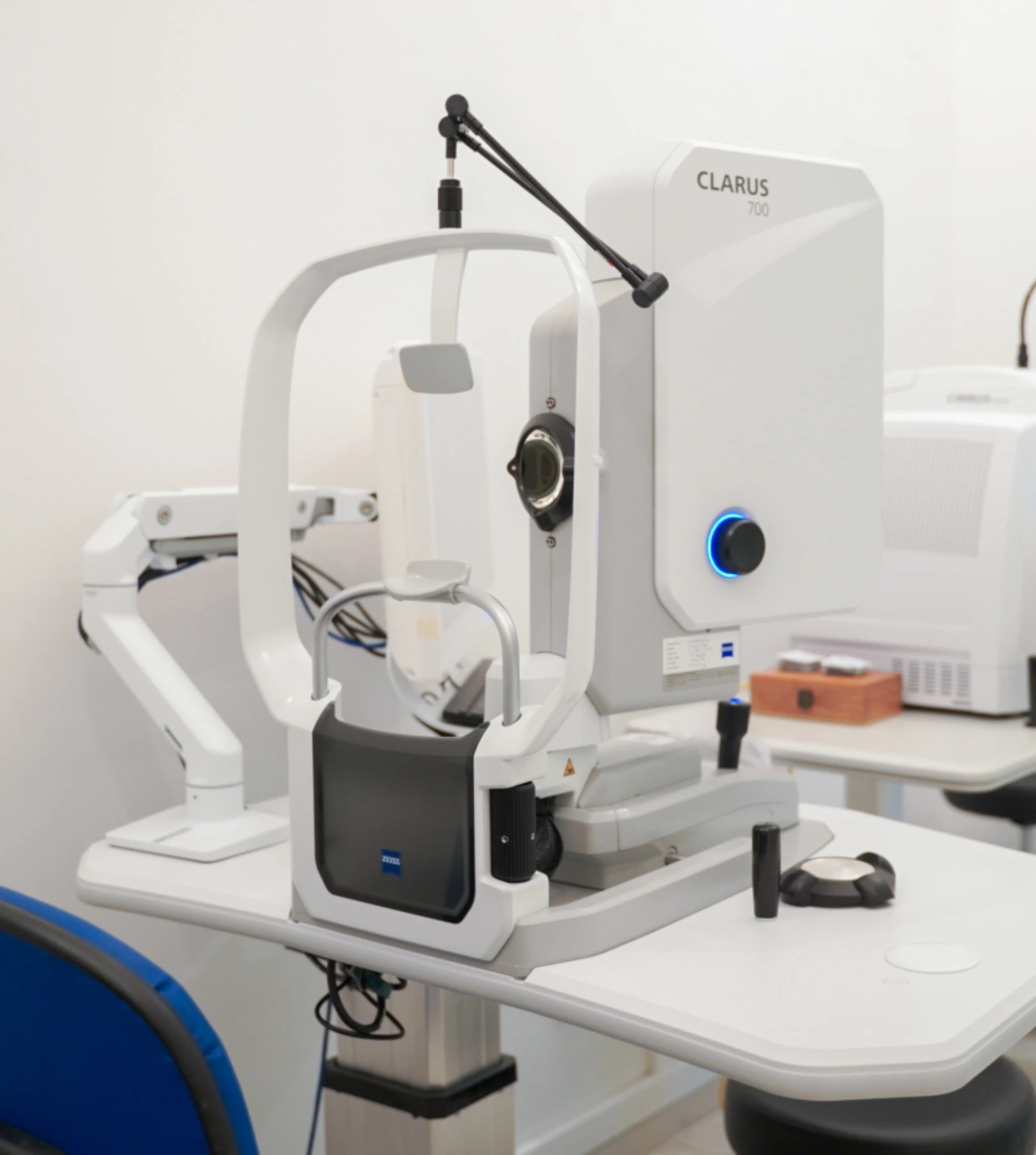
We use a combination of detailed eye examination and advanced retinal imaging to assess the macula, retina, optic nerve, and retinal blood vessels. These tests allow us to identify structural and functional changes, determine the type and severity of the condition, and establish a clear baseline for ongoing monitoring and treatment planning.
Measures how clearly you can see and helps identify any reduction in central vision.
Documents the size, location, colour and appearance of the choroidal naevus so that changes can be compared over time.
Highlights changes in the retinal pigment epithelium, a cellular layer under the naevus, and can demonstrate areas of increased autofluorescence associated with lipofuscin (waste product of retinal activity) or orange pigments over the lesion.
Assesses the retinal circulation and leakage around the lesion, helping identify associated retinal changes and providing additional information when distinguishing between different choroidal lesions.
Provides deeper imaging of the choroidal circulation and can help assess the vascular characteristics of selected choroidal lesions.
Measures the thickness and internal characteristics of the lesion, particularly when assessing larger or elevated lesions and monitoring for changes over time.
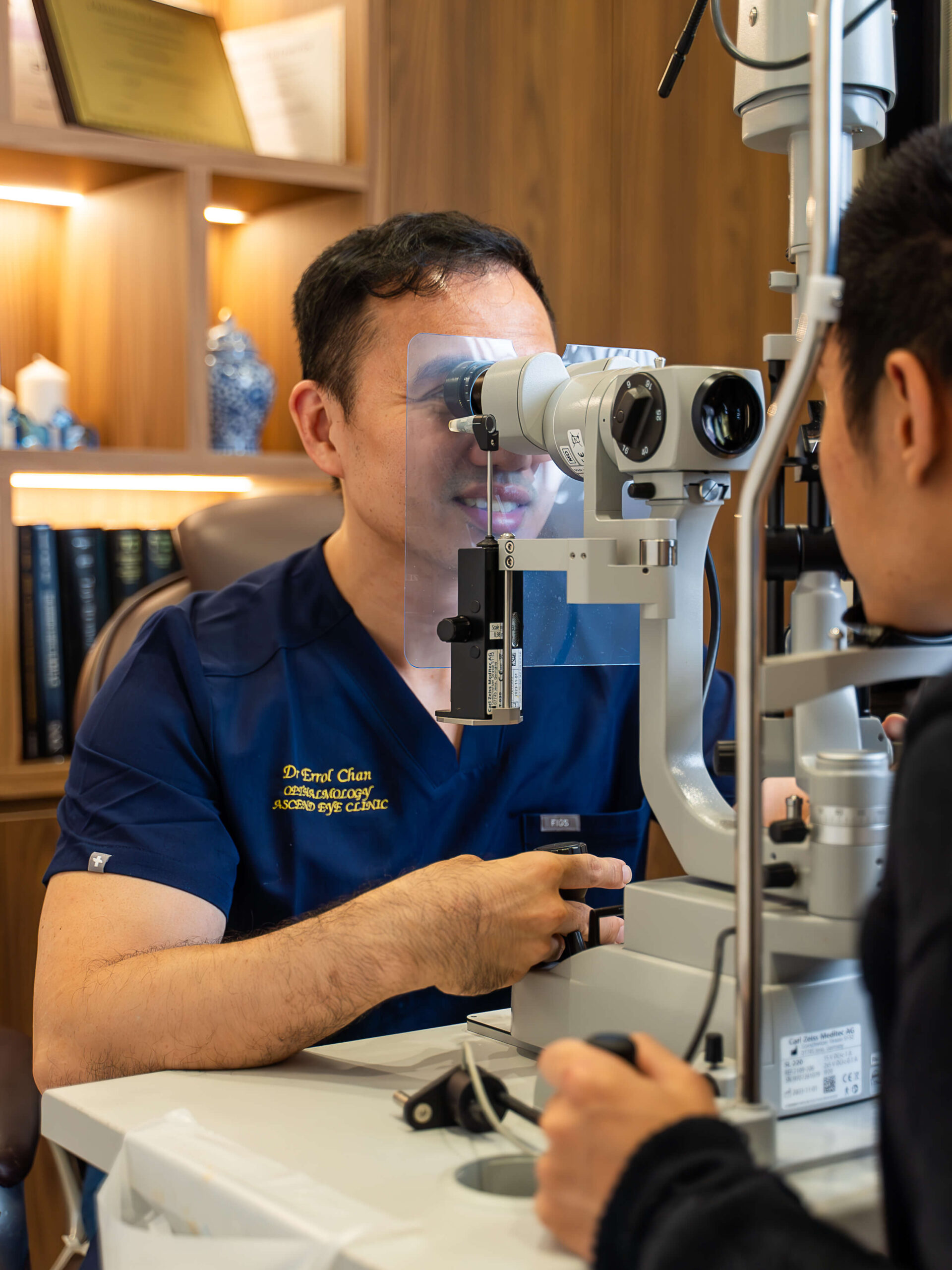
The vast majority of pigmented choroidal lesions are choroidal naevi, which are benign and do not require treatment. They are monitored with regular dilated retinal examinations and specialised retinal imaging to document their appearance and check for changes in size, thickness or in the surrounding retina.
Choroidal naevi should be monitored at regular intervals. Our eye specialists will discuss with you regarding the frequency of follow-up required.
Naevi with structural features that raise concern for malignancy or possible transformation may require closer and more frequent monitoring.
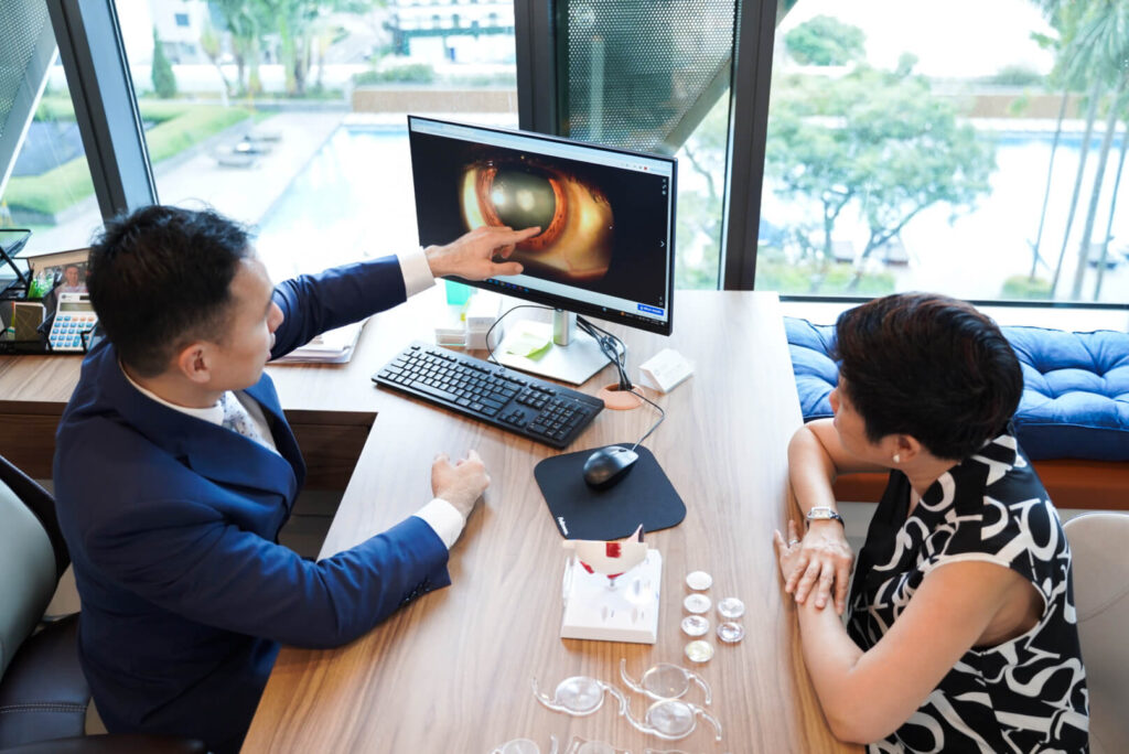
Your consultation begins with a detailed assessment of your vision and eye health. We will review any visual symptoms, your medical and ocular history, and any previous eye examinations or imaging.
We will also discuss whether the choroidal naevus was identified recently or has been monitored previously, including any known changes over time.
A detailed retinal examination and imaging are performed to assess the choroidal naevus and document its appearance. This may include widefield fundus photography, optical coherence tomography and fundus autofluorescence.
These investigations allow us to assess features such as the size, location and surface characteristics of the naevus, and to establish a baseline for comparison at future visits.
Most choroidal naevi remain stable and do not require treatment. Monitoring focuses on identifying any change in size or appearance over time and detecting features that require closer assessment.
Your follow-up schedule will be tailored according to the appearance of the naevus, retinal findings and previous imaging. Repeat retinal photography and imaging will allow direct comparison over time so that any meaningful changes are identified promptly.
Choroidal naevi and tumours are often detected during a dilated retinal examination. Most are benign, but appropriate imaging may be needed to document their appearance and determine whether monitoring or treatment is required.
Book a consultation at Ascend Eye Clinic for a comprehensive retinal examination and retinal imaging. We will explain the findings clearly and advise you on the recommended follow-up.
In most cases, no. A choroidal naevus is a benign collection of pigment-producing cells inside the eye. Most remain stable and never cause problems, although monitoring is important to check for any changes over time.
No. A choroidal naevus is benign. It is similar to having a freckle or mole within the eye.
No. The word “tumour” simply refers to a growth or lesion. Choroidal tumours can be benign, such as a choroidal naevus, or malignant, such as a choroidal melanoma.
Most choroidal naevi remain benign throughout life. A small number develop changes that raise concern for melanoma, which is why certain lesions are monitored with retinal imaging.
We assess characteristics such as the lesion’s size, thickness, shape and appearance, as well as whether there is fluid or other changes in the surrounding retina. Comparing imaging over time is particularly useful for identifying growth.
Many choroidal naevi remain unchanged. Some can show small changes without being malignant, while progressive or suspicious growth requires further assessment.
Not always. A choroidal lesion can change without causing noticeable symptoms, which is why examination and retinal imaging are important even when your vision feels normal.
Most do not affect vision. Visual changes are more likely when the lesion is close to the macula or causes fluid or changes in the overlying retina.
Follow-up allows us to document that the lesion remains stable. Retinal photographs and other imaging provide an objective comparison between visits and help identify any concerning changes early.
This depends on the appearance of the lesion and whether previous imaging has shown any change. A stable, low-risk naevus generally requires less frequent monitoring than a lesion with suspicious features.
No. A stable choroidal naevus that is not causing problems does not require removal or treatment and is usually monitored instead.
We will carry out further assessment and imaging to determine the nature of the lesion. If choroidal melanoma is suspected or confirmed, treatment is planned according to its size, location and effect on the eye.
Treatment commonly involves radiotherapy, including plaque brachytherapy, where a radioactive plaque is temporarily positioned on the outside of the eye next to the tumour. Surgery is used in selected cases depending on the size and location of the melanoma.
Yes. Unlike a choroidal naevus, choroidal melanoma is malignant and can spread outside the eye. Patients diagnosed with melanoma therefore require appropriate ocular treatment and further assessment as part of their ongoing care.
Arrange an earlier assessment if you develop new or increasing blurred vision, distorted or wavy vision, flashes of light, floaters, or a shadow or blurred area in your vision.
Hi, I would like to book a consultation with Dr Errol Chan from Ascend Eye Clinic
