
When vision changes after cataract surgery, timely assessment helps identify the cause and determine the right treatment. We provide careful evaluation of dislocated intraocular lenses and personalised treatment options to restore stability and improve visual function.
Cataract surgery is a commonly performed procedure that replaces the eye’s cloudy natural lens with a clear, artificial lens, known as an intraocular lens or IOL.
An IOL dislocation occurs when the artificial lens shifts away from its intended position because the structures supporting it have weakened or become damaged. This can either occur shortly after cataract surgery or develop many years later.
The risk of a dislocated intraocular lens is higher in people with:
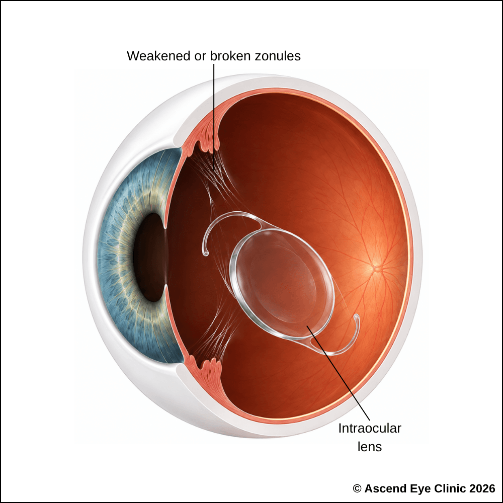



The main symptom of a dislocated intraocular lens is a change in vision. The extent of the visual disturbance depends on how far the lens has shifted.
A significantly dislocated or dropped IOL can affect other structures within the eye, resulting in inflammation, raised eye pressure, corneal swelling, or retinal complications. Prompt assessment is important when vision changes suddenly after cataract surgery.
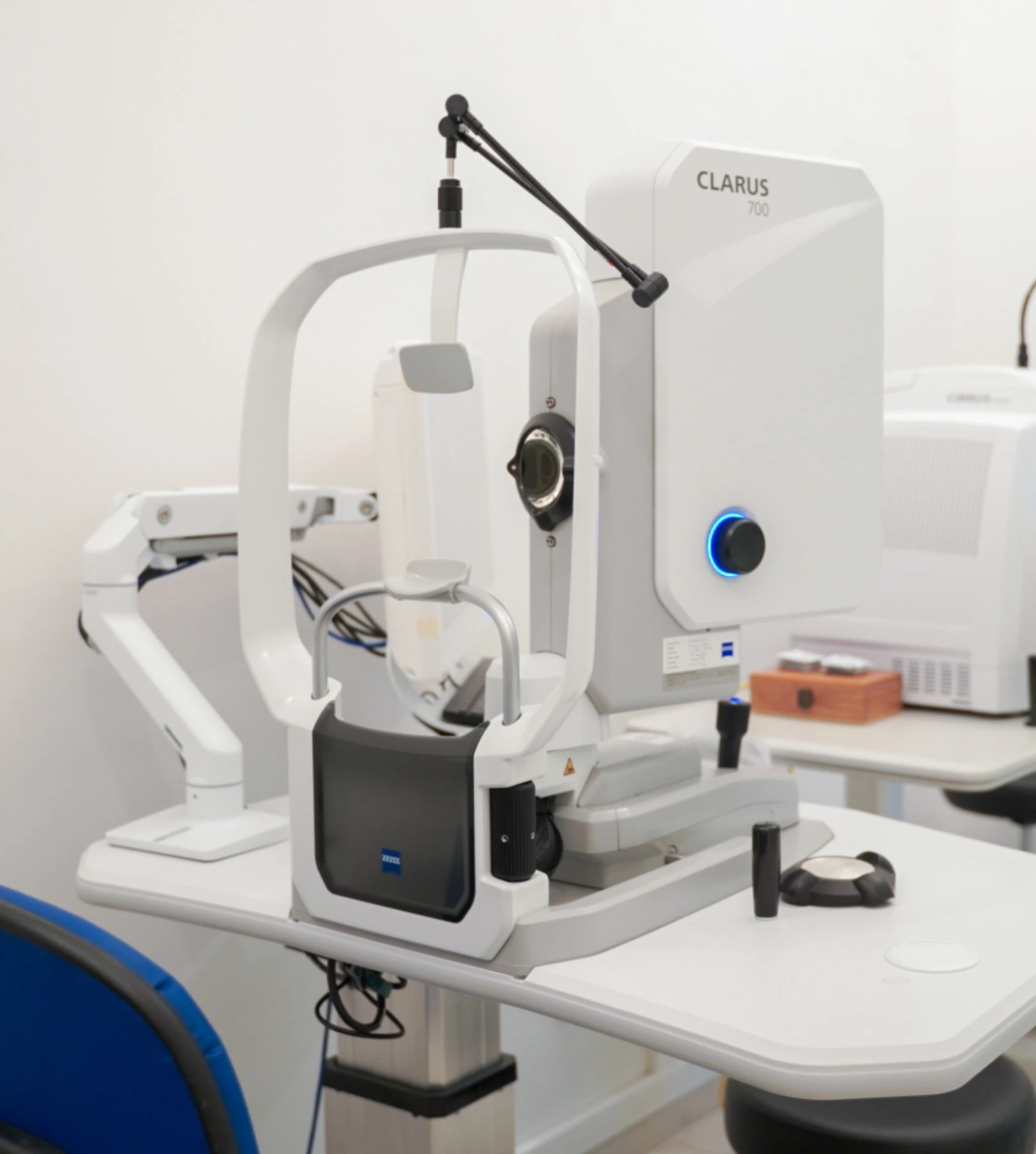
We use a combination of detailed eye examination and advanced retinal imaging to assess the macula, retina, optic nerve, and retinal blood vessels. These tests allow us to identify structural and functional changes, determine the type and severity of the condition, and establish a clear baseline for ongoing monitoring and treatment planning.
Measures how clearly you can see and helps identify any reduction in central vision.
Measures your refractive error and assesses changes caused by movement or tilting of the intraocular lens.
Examines the position and stability of the intraocular lens and assesses the cornea, anterior chamber, iris and other structures at the front of the eye for any associated changes.
Captures detailed images of the retina and optic nerve and helps assess the posterior segment, particularly when the intraocular lens has displaced backwards into the eye.
Provides cross-sectional images of the retina to assess the macula for swelling or other retinal changes that may affect vision.
Measures the dimensions of the eye and calculates the appropriate intraocular lens power if IOL exchange or replacement is planned.
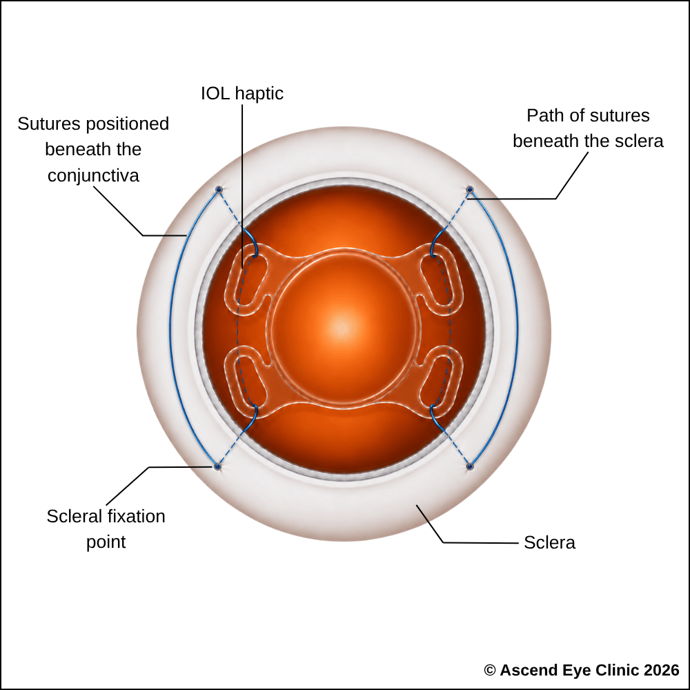
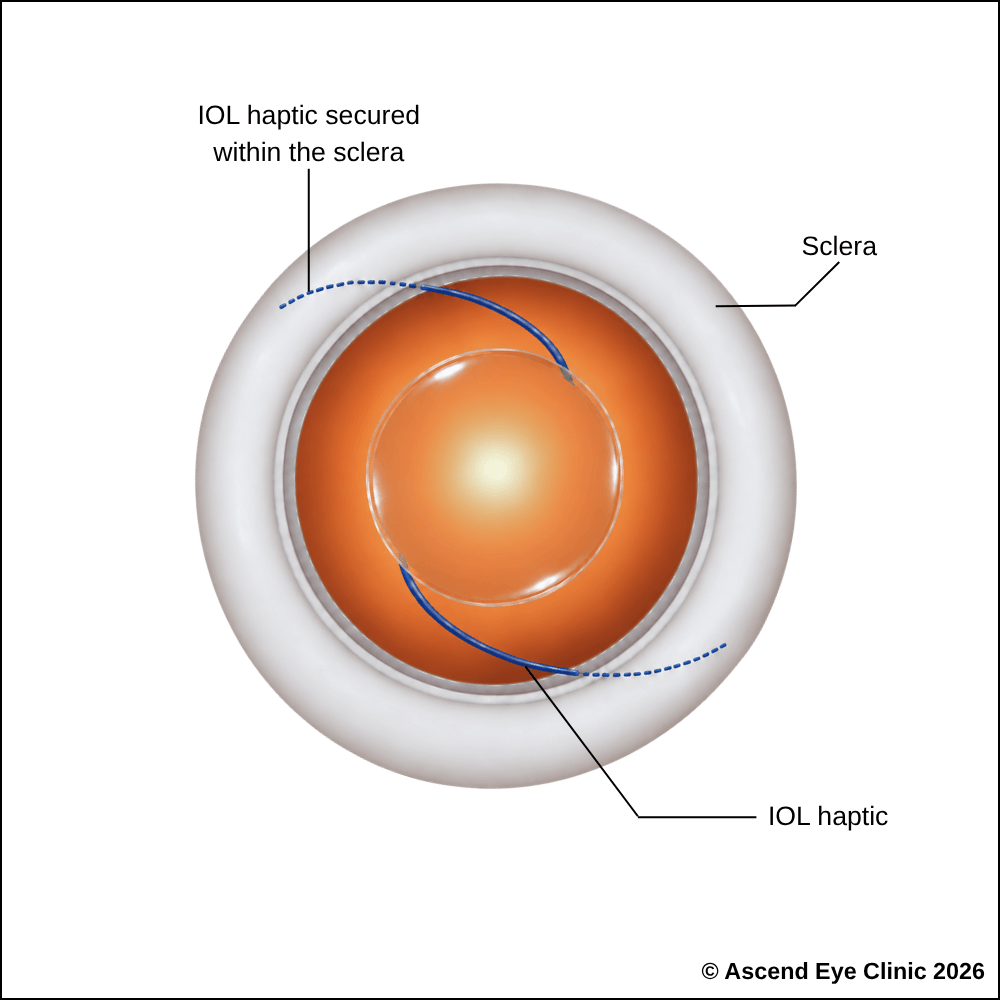
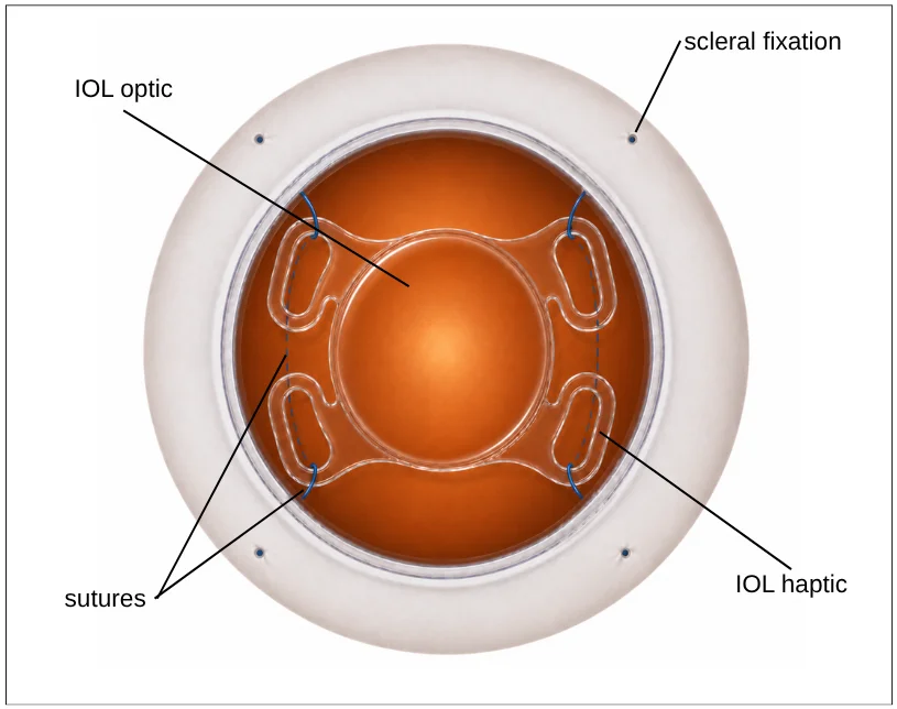
Before the replacement IOL is positioned, a support structure is created in the sclera to secure the new lens without relying on the natural lens capsule. The haptics, or “legs” of the IOL, are used as part of this fixation, and any external fixation points are concealed beneath the conjunctiva.
In IOL exchange surgery, an unstable or displaced IOL is removed and replaced with a new lens. The replacement IOL is positioned behind the iris and fixed to the sclera, the white outer wall of the eye.
The replacement IOL is secured using either the Yamane sutureless technique, where the haptics of the lens are passed through the sclera and secured without sutures, or four-point Gore-Tex® suture fixation, where the lens is attached to the sclera at four points using Gore-Tex® sutures. Both techniques provide stable support for the replacement IOL without relying on the natural lens capsule.
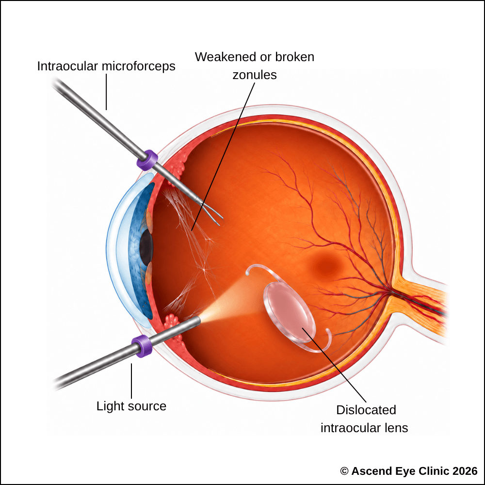
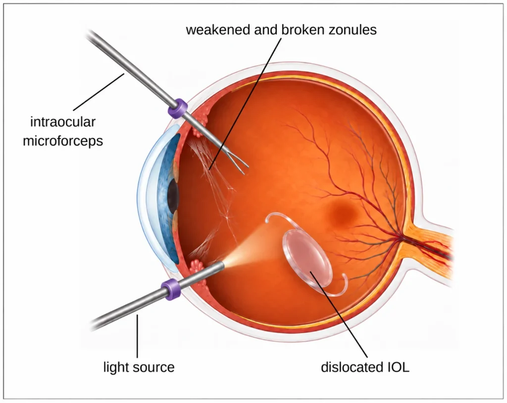
If the displaced IOL has fallen into the vitreous cavity at the back of the eye, a vitrectomy is required to safely retrieve it before the above procedure is performed. This is a microsurgical keyhole procedure performed through several small ports in the white part of the eye.
Fine instruments are used to remove the vitreous gel and free the displaced IOL. The IOL is then brought forward from the back of the eye into the front of the eye, where it is removed through an incision. In some cases, the IOL needs to be cut into smaller pieces before it can be safely removed. The retina and surrounding structures are then inspected to ensure the back of the eye remains stable.
A secondary IOL is then implanted and secured to the sclera using either the Yamane sutureless technique or four-point Gore-Tex® suture fixation, as described above.
Surgery is recommended when the IOL affects vision, becomes unstable or drops into the back of the eye. After assessing the amount of support remaining within the eye, we will recommend one of two approaches.
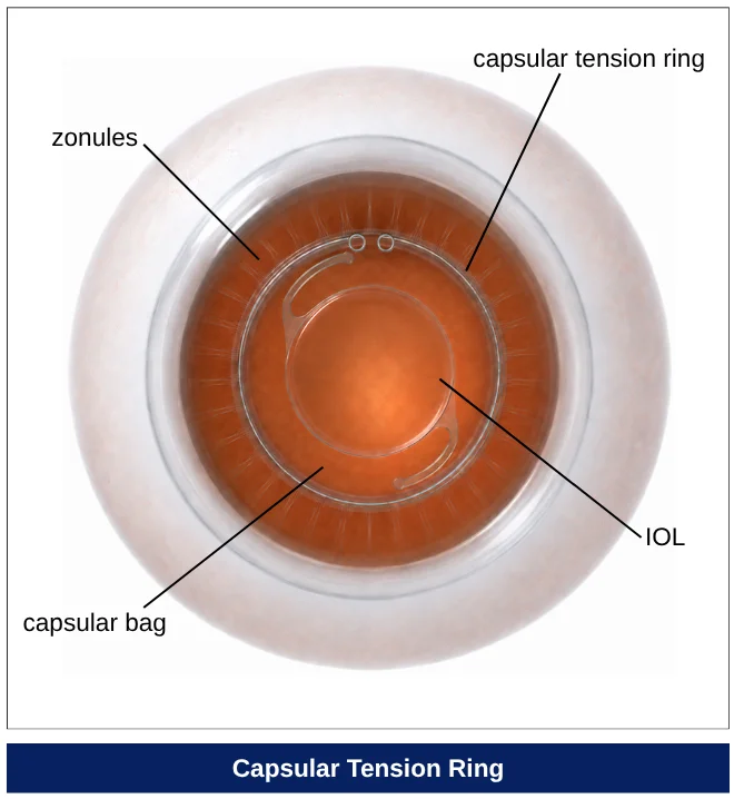
When the IOL remains within the capsular bag and there is enough remaining support, we can preserve the existing lens rather than replace it. A capsular tension ring (CTR) is inserted into the weakened capsular bag to redistribute support around the lens, helping to recentre the IOL and keep it securely positioned.
CTR insertion is an outpatient procedure performed in an operating theatre, usually under local anaesthesia with sedation. Through a small incision at the front of the eye, fine instruments are used to access the capsular bag and carefully reposition the existing IOL. The capsular tension ring is then inserted around the inside of the bag, where it expands gently against the capsule to provide more even support and improve the stability of the IOL.
The small incision is self-sealing, and patients can return home on the same day after a short period of observation. Vision is usually blurred initially and gradually improves as the eye heals and the IOL settles into its new position.
If an IOL becomes too unstable or there is no longer enough support to hold it securely in place, the lens may need to be removed and replaced.
The surgical approach depends on where the displaced lens is sitting within the eye. If the lens is still positioned near the pupil and can be reached safely from the front of the eye, it can usually be brought forward through the pupil and removed without the need for a vitrectomy. A new secondary lens implant can then be placed and secured to the sclera, the strong white wall of the eye.
If the lens has dropped further back into the vitreous cavity, a vitrectomy is required first. During vitrectomy, the vitreous gel surrounding the displaced lens is carefully removed to free the IOL, create space for the instruments to work and minimise traction on the retina. The lens can then be safely brought forward and removed before a new secondary IOL is implanted.
The replacement lens is secured in a stable position behind the iris using either Gore-Tex suture scleral fixation or the Yamane sutureless scleral fixation technique. The choice of technique depends on the individual eye and which method is most likely to provide a stable, well-centred lens position.
Vitrectomy is a keyhole procedure performed in an operating theatre under general anaesthesia. Fine microsurgical instruments are introduced through tiny ports in the white wall of the eye, allowing the safe removal of the vitreous gel and retrieve the displaced lens when it has fallen into the back of the eye.
Vision is usually blurred initially after surgery and gradually improves as the eye heals and the new IOL settles into position. The extent of visual recovery depends on factors such as the health of the retina, macula and other structures within the eye.
From your initial consultation to treatment and follow-up care, our team will guide you through every stage of your journey. We will take the time to explain your condition, answer your questions and help you feel informed and supported throughout your care.

Your consultation begins with a detailed assessment of your vision and eye health. Depending on your condition, this may include measurements of your vision and eye pressure, examination of the front and back of the eye, and advanced diagnostic imaging.
Our team will guide you through each test and explain what is being assessed. Your results will then be carefully reviewed to understand your individual needs and determine whether monitoring or treatment may be recommended.
Every eye and every patient is different. Your treatment plan will be tailored according to your examination findings, visual needs, general health and lifestyle.
We will explain the recommended treatment, its expected benefits, possible risks and any suitable alternatives in a clear and straightforward manner. You will have time to ask questions and consider your options before deciding how you would like to proceed.
Before your treatment, our team will guide you through the necessary preparations, paperwork and medication instructions. Where applicable, we will explain how to use your prescribed eye drops and how to care for your eyes afterwards.
We understand that undergoing a procedure can feel unfamiliar or worrying. Our team will be available to answer your questions and ensure that you know what to expect before the day of your treatment.
On the day of your procedure, you will arrive at the hospital or surgical centre where your operation has been scheduled. The surgical team will help you prepare and explain what will happen at each stage.
Anaesthetic eye drops or other forms of anaesthesia may be used, depending on the procedure, to keep you comfortable. Your doctor and the surgical team will support and reassure you throughout the operation.
Most eye surgeries are performed as day procedures, although the duration and recovery process will depend on the treatment you receive. Your vision may be temporarily blurred afterwards, so you should arrange for someone to accompany you home and should not drive unless your doctor has advised that it is safe to do so.
We will provide clear instructions on how to care for your eyes, use any prescribed medication and manage your activities during recovery. We will also explain which symptoms are expected and when you should contact the clinic for further advice.
Follow-up appointments allow us to monitor your vision, assess your recovery and identify any concerns at an early stage. Our team remains accessible should you have any questions or concerns throughout your recovery.
A displaced or unstable lens implant can cause blurred, distorted or double vision. At Ascend Eye Clinic, we will assess the position of your IOL and recommend the most suitable approach to restore stable lens support, including scleral-fixated intraocular lens surgery where required.
Book a consultation with Ascend Eye Clinic for a detailed evaluation and personalised treatment plan.
A displaced IOL has shifted away from its intended position inside the eye but may still remain partly supported by the capsular bag or surrounding structures. A dropped IOL has fallen further backwards into the vitreous cavity, which is the gel-filled space at the back of the eye. A dropped IOL usually requires retinal surgery to safely retrieve or reposition the lens.
An IOL may dislocate many years after otherwise successful cataract surgery because the fine fibres, known as zonules, that support the capsular bag can gradually weaken. As this support becomes less stable, the entire capsular bag and IOL may shift or become loose. Certain eye conditions, previous surgery or trauma can accelerate this process.
IOL dislocation is more likely when the structures supporting the lens are weakened. Risk factors include previous eye trauma, retinal or vitreous surgery, uveitis, pseudoexfoliation syndrome, high myopia and certain connective tissue disorders. The risk can also increase over time following cataract surgery as the supporting zonules naturally become weaker.
Symptoms depend on how far the IOL has shifted. Patients may notice blurred or fluctuating vision, double vision, glare, visual distortion or difficulty focusing. Some may notice the edge of the lens implant or experience a sudden change in their glasses prescription. Symptoms can sometimes change with head position if the lens is unstable.
A dislocated IOL itself is usually painless. However, if the displaced lens causes inflammation, increased eye pressure, corneal problems or other complications, the eye may become uncomfortable or painful. New pain, redness or a sudden deterioration in vision should therefore be assessed promptly.
Not every displaced IOL requires immediate surgery. If the lens has only moved slightly, remains stable and does not significantly affect vision or the health of the eye, it may be monitored with regular eye examinations. Surgery may become necessary if the position changes, vision worsens or complications develop.
Surgery is usually considered when the IOL has moved enough to affect vision, becomes unstable or causes other problems within the eye. Treatment may also be required if the IOL drops into the vitreous cavity. Depending on the condition of the existing lens and its supporting structures, the IOL may be repositioned, stabilised or replaced with a different lens.
Your eye surgeon will perform a detailed examination to assess the position and stability of the IOL. The pupil is usually dilated so that the lens, capsular bag, retina and other structures can be examined properly. Additional imaging may be used when necessary to assess the retina, macula or other parts of the eye and to help plan treatment.
In some cases, the existing IOL and capsular bag can be preserved rather than replaced. Capsular bag stabilisation involves supporting and recentring the capsular bag–IOL complex when sufficient capsule remains. Specialised devices such as a capsular tension ring or segment may be used, sometimes together with fixation to the wall of the eye, to improve stability.
A scleral-fixated intraocular lens (SFIOL) is an IOL that is secured to the sclera, which is the strong white wall of the eye. This technique is used when the natural capsular bag no longer provides enough support to hold an IOL safely in position. Different fixation methods can be used depending on the individual eye and the type of lens selected.
A vitrectomy may be required when the IOL or part of the capsular bag has moved into the vitreous cavity at the back of the eye. It may also be needed when vitreous gel has moved around or in front of the displaced lens. During vitrectomy, the vitreous is carefully removed so that the IOL can be safely retrieved, repositioned or exchanged while protecting the retina.
This depends on the condition of the IOL, how far it has moved and whether there is enough support remaining inside the eye. In some cases, the existing lens can be recentred and secured. In others, it may be safer to remove it and implant a new lens in a more stable position.
The surgical approach depends on where the IOL has moved, the amount of remaining capsular support and whether the vitreous or retina is involved. We will assess these factors during the eye examination and recommend the technique that provides the safest and most stable lens position for your eye.
The aim of surgery is to restore a stable lens position and improve optical focus. Many patients experience a significant improvement in vision once the IOL is properly positioned. However, the final visual outcome also depends on the health of other parts of the eye, including the cornea, macula, retina and optic nerve, as well as any pre-existing eye conditions.
Re-dislocation is uncommon when the IOL has been securely repositioned or fixated, although no surgical technique can completely eliminate the possibility of future movement. Regular follow-up allows your eye surgeon to monitor the position of the lens, eye pressure, retina and surrounding structures and identify any changes early.
Hi, I would like to book a consultation with Dr Errol Chan from Ascend Eye Clinic
