
A retinal tear can develop suddenly and may progress to a retinal detachment if left untreated. Prompt assessment allows us to identify the tear early, examine the surrounding retina carefully, and provide timely treatment to seal the affected area before it threatens your vision.
The retina is the thin, light-sensitive layer at the back of the eye that allows us to see. A retinal tear develops when the vitreous, the clear gel that fills the eye, pulls strongly enough on the retina to create a small break.
As we age, the vitreous naturally becomes more liquid and gradually separates from the retina. This is known as posterior vitreous detachment, or PVD, and usually occurs without causing any problems. However, if the vitreous remains firmly attached to part of the retina as it separates, the pulling force may cause the retina to tear. Retinal tears may also occur following an injury to the eye.
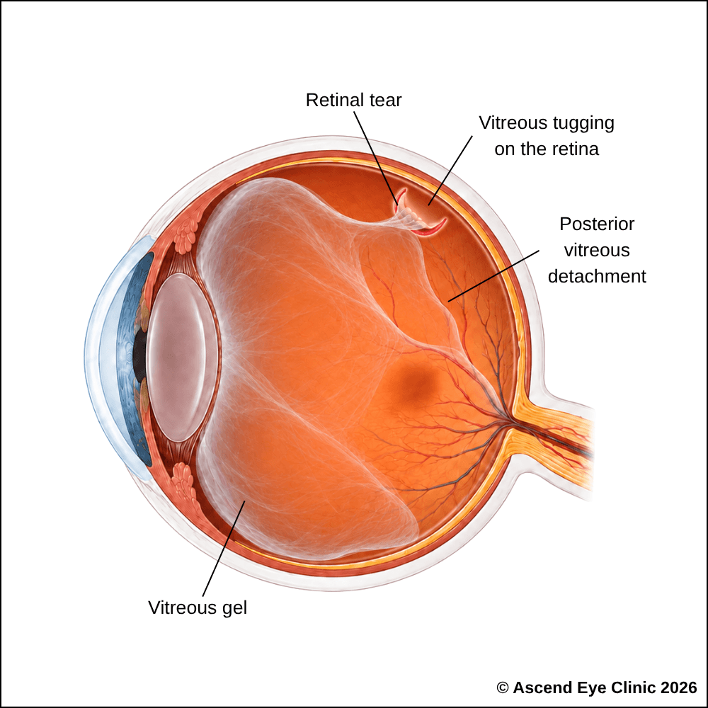
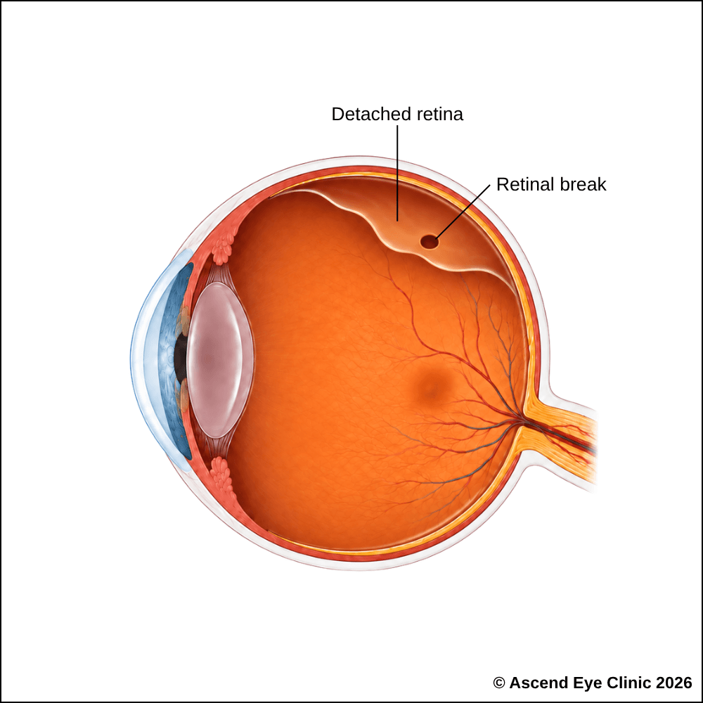
If fluid passes through the tear and collects beneath the retina, it may cause the retina to separate from the back of the eye. This is known as a retinal detachment and can result in permanent vision loss if treatment is delayed. When identified early, a retinal tear can often be sealed with laser treatment to reduce the risk of detachment.

Although retinal tears can occur without any obvious risk factors, they are more common in people who:



Retinal tears are usually painless. Warning signs include a sudden increase in floaters (dark spots or lines in your vision) and new flashes of light at the side of your vision.
A shadow or curtain may indicate that the retina has started to detach and requires urgent assessment. Early detection allows treatment to be performed before more serious vision loss develops.
We use a combination of detailed eye examination and advanced retinal imaging to assess the macula, retina, optic nerve, and retinal blood vessels. These tests allow us to identify structural and functional changes, determine the type and severity of the condition, and establish a clear baseline for ongoing monitoring and treatment planning.
Measures how clearly you can see and helps identify any reduction in central vision.
Provides a magnified examination of the eye using a specialised lens to assess the peripheral retina, identify retinal tears or holes, and check for signs of retinal detachment.
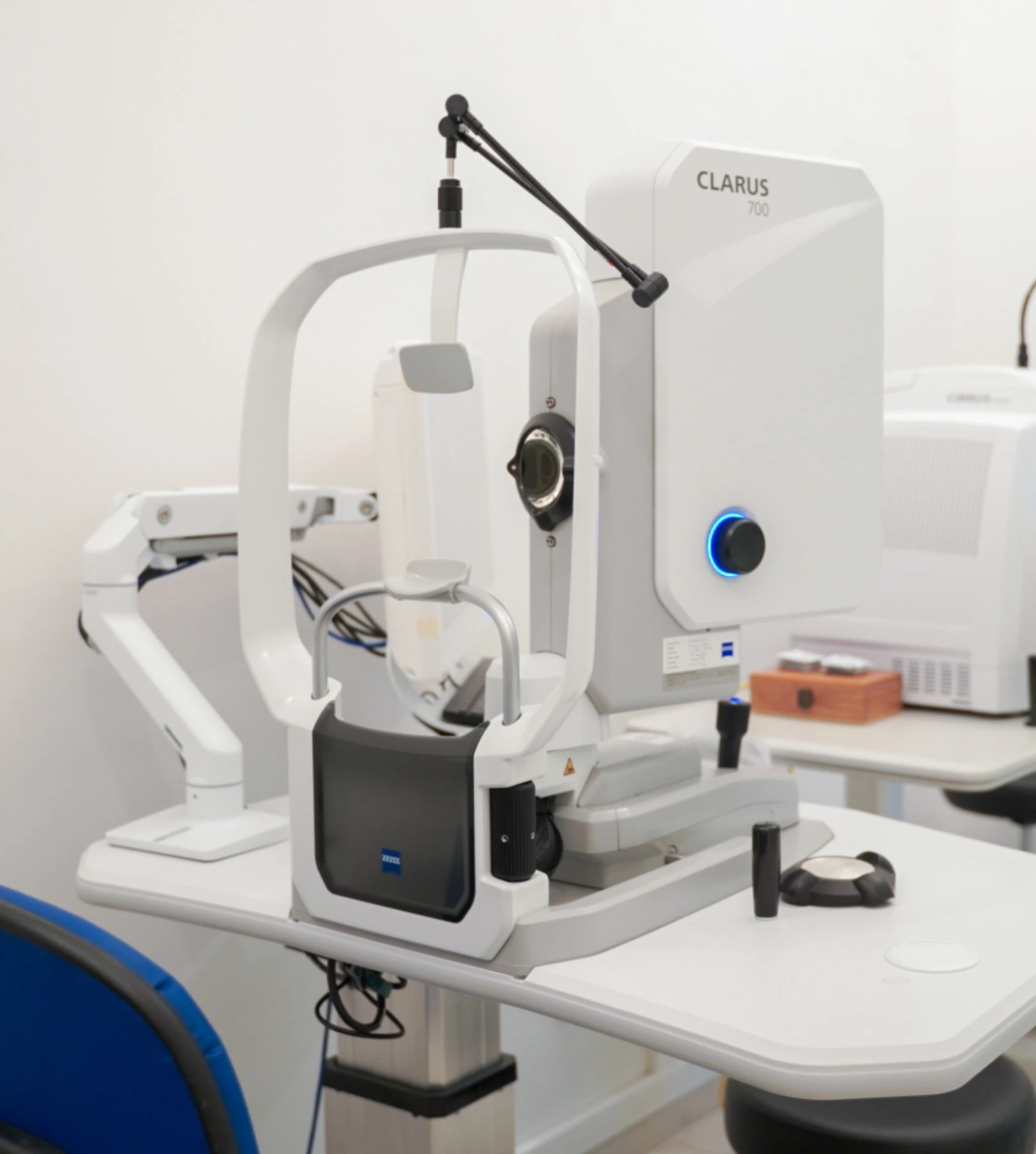
Captures detailed images of the peripheral retina to identify retinal tears, holes, areas of retinal detachment and other associated abnormalities.
Provides cross-sectional images of the retina to assess the macula and individual retinal layers for swelling, fluid or other structural changes.
Retinal tears require prompt treatment to seal the tear and reduce the risk of fluid passing underneath the retina, which can lead to a retinal detachment. Early treatment helps stabilise the retina and prevent the condition from progressing.
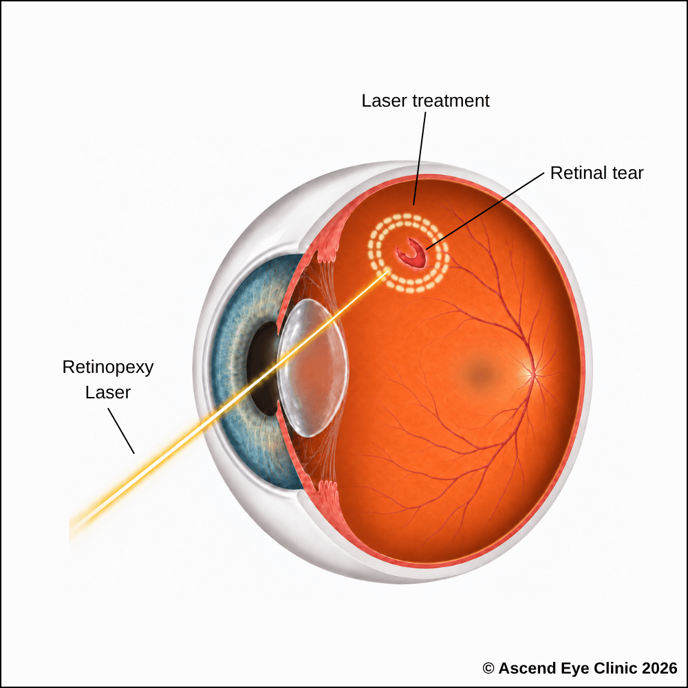
Laser retinopexy uses a laser to place a series of small, controlled spots around a retinal tear or hole. As these areas heal, they form a firm adhesion between the retina and the underlying tissue, creating a protective barrier around the break and reducing the risk of fluid passing underneath the retina and causing a retinal detachment.
Laser retinopexy is performed as an outpatient procedure with no hospitalisation required. Before treatment, numbing eye drops are applied and a special contact lens is placed on the eye to provide a clear view of the retinal tear and allow the laser to be delivered precisely. Several rows of laser spots are then applied around the affected area to secure the retina. You will see bright flashes of light during the procedure, but treatment is generally well tolerated with little or no pain.
After the procedure, vision will be temporarily blurred due to the bright laser light and dilating eye drops used during treatment. This will gradually clear over the following hours as the effects wear off. The laser adhesion strengthens as the treated area heals over the following days. Follow-up examinations are important to assess healing and confirm that the retinal tear is adequately secured by the laser treatment.
From your initial consultation to treatment and follow-up care, our team will guide you through every stage of your journey. We will take the time to explain your condition, answer your questions and help you feel informed and supported throughout your care.

Your consultation begins with a detailed assessment of your vision and eye health. Depending on your condition, this may include measurements of your vision and eye pressure, examination of the front and back of the eye, and advanced diagnostic imaging.
Our team will guide you through each test and explain what is being assessed. Your results will then be carefully reviewed to understand your individual needs and determine whether monitoring or treatment may be recommended.
Every eye and every patient is different. Your treatment plan will be tailored according to your examination findings, visual needs, general health and lifestyle.
We will explain the recommended treatment, its expected benefits, possible risks and any suitable alternatives in a clear and straightforward manner. You will have time to ask questions and consider your options before deciding how you would like to proceed.
Before your treatment, our team will guide you through the necessary preparations, paperwork and medication instructions. Where applicable, we will explain how to use your prescribed eye drops and how to care for your eyes afterwards.
We understand that undergoing a procedure can feel unfamiliar or worrying. Our team will be available to answer your questions and ensure that you know what to expect before the day of your treatment.
On the day of your procedure, you will arrive at the hospital or surgical centre where your operation has been scheduled. The surgical team will help you prepare and explain what will happen at each stage.
Anaesthetic eye drops or other forms of anaesthesia may be used, depending on the procedure, to keep you comfortable. Your doctor and the surgical team will support and reassure you throughout the operation.
Most eye surgeries are performed as day procedures, although the duration and recovery process will depend on the treatment you receive. Your vision may be temporarily blurred afterwards, so you should arrange for someone to accompany you home and should not drive unless your doctor has advised that it is safe to do so.
We will provide clear instructions on how to care for your eyes, use any prescribed medication and manage your activities during recovery. We will also explain which symptoms are expected and when you should contact the clinic for further advice.
Follow-up appointments allow us to monitor your vision, assess your recovery and identify any concerns at an early stage. Our team remains accessible should you have any questions or concerns throughout your recovery.
A retinal tear may progress to retinal detachment if fluid begins to collect beneath the retina. Prompt assessment allows the retina to be examined carefully and, where necessary, treated before this occurs.
Book a retinal consultation at Ascend Eye Clinic for a detailed examination and personalised advice on whether laser treatment or monitoring is recommended.
A retinal tear is a break in the retina, the light-sensitive layer lining the back of the eye. It most commonly occurs when the vitreous gel inside the eye pulls firmly on the retina as it separates with age. A tear creates an opening through which fluid can pass beneath the retina, increasing the risk of retinal detachment.
A retinal tear commonly causes a sudden increase in floaters, new flashes of light, or both. Some people describe many small black dots, cobweb-like shapes or streaks appearing unexpectedly in their vision. If the retina begins to detach, a dark shadow or curtain can also develop in the visual field.
No. Some retinal tears cause no noticeable symptoms and are discovered during a dilated retinal examination. This is why a detailed examination of the peripheral retina is important, particularly in people with risk factors such as high eye power, previous retinal tears or recent eye trauma.
Yes. Most retinal tears begin in the peripheral retina, so central vision can remain completely clear at first. A person can therefore have a significant retinal tear while still reading normally and seeing clearly straight ahead. Central vision becomes threatened if a retinal detachment progresses towards the macula.
You should arrange an urgent retinal assessment. Sudden new floaters or flashes can occur when the vitreous separates from the retina, and this process can produce a retinal tear. We examine the peripheral retina carefully to determine whether a tear is present and whether treatment is required.
The most common cause is posterior vitreous detachment, where the vitreous gel naturally separates from the retina. If the vitreous remains firmly attached at a particular area, it can pull strongly enough to tear the retina. Eye trauma and certain retinal conditions can also cause retinal tears.
Yes. People with higher degrees of short-sightedness generally have longer eyes and thinner, more stretched peripheral retinas. This increases the likelihood of peripheral retinal degeneration, retinal tears and retinal detachment. Regular retinal examinations are particularly important when the eye power is high.
Yes. A significant impact to the eye or head can create sudden traction on the retina and produce a retinal tear or detachment. New floaters, flashes, blurred vision or a shadow in the vision following trauma should be assessed promptly.
Yes. Retinal tears and detachments can occur after cataract surgery, particularly in eyes that are highly short-sighted or already have peripheral retinal abnormalities. The overall risk remains low, but new floaters, flashes or a shadow in the vision after eye surgery require prompt retinal assessment.
We dilate the pupil and examine the retina carefully, including the far peripheral retina where most tears occur. Retinal imaging can provide additional information, but a thorough dilated examination remains essential because very peripheral tears are not always captured on standard retinal scans.
A retinal tear should not be assumed to heal by itself. When a tear has features that increase the risk of retinal detachment, we seal it with retinal laser or, in selected cases, cryotherapy. Some low-risk retinal breaks can be safely monitored when examination shows that treatment is not necessary.
Fluid from inside the eye can pass through the tear and collect underneath the retina. As more fluid accumulates, the retina can separate from the underlying eye wall, producing a retinal detachment. Treating an appropriate retinal tear before this happens is considerably simpler than treating an established retinal detachment.
No. The risk depends on the type, size and location of the tear, whether the vitreous is continuing to pull on it, and whether fluid has already started passing underneath the retina. We assess these features carefully to determine which tears require treatment and which can be monitored safely.
Most retinal tears requiring treatment are sealed with a retinal laser. The laser creates a series of controlled spots around the tear, producing a firm adhesion between the retina and the underlying tissue. This forms a protective barrier that reduces the chance of fluid passing through the tear and progressing to a retinal detachment. Cryotherapy can be used in selected cases when laser treatment is not suitable.
Anaesthetic drops are used to numb the eye before treatment. A special contact lens is then placed on the eye so we can accurately direct the laser around the retinal tear. You will see bright flashes of light and can feel some pressure or brief discomfort, but the treatment is generally well tolerated and is completed as an outpatient procedure.
The laser begins creating an adhesion around the tear soon after treatment, with the scar becoming progressively stronger over the following days. A firm protective seal generally develops over approximately one to two weeks. We arrange follow-up examinations to confirm that the laser has adequately surrounded the tear.
Laser treatment seals the retinal tear, but it does not remove the vitreous changes that originally caused the floaters and flashes. Flashes usually reduce as vitreous traction settles. Floaters often become less noticeable with time as they move away from the central visual axis and the brain adapts to them.
Normal light activities can usually continue after retinal laser treatment. We generally advise avoiding strenuous exercise, heavy lifting and high-impact activities for a short period while the laser adhesion strengthens. Flying is not restricted after laser treatment alone; however, flying is strictly prohibited if a gas bubble has been placed inside the eye until the gas has completely disappeared.
Yes. Laser seals the specific tear that has been treated, but it does not prevent the vitreous from pulling on another part of the retina. A new tear can therefore develop elsewhere. Any new burst of floaters, renewed flashes or a curtain or shadow in the vision requires another urgent retinal examination.
Yes. We review the retina after treatment to confirm that the tear is securely surrounded and to check for additional retinal breaks or early detachment. Further follow-up depends on the appearance of the retina, your eye power, the condition of the vitreous and whether there are other areas of peripheral retinal weakness.
Hi, I would like to book a consultation with Dr Errol Chan from Ascend Eye Clinic
