
Excessive tearing can be uncomfortable, disruptive and may have several different causes, including dry eye, eyelid problems or blockage of the tear drainage system. A thorough eye assessment helps identify the underlying cause so that treatment can be tailored appropriately and symptoms managed effectively.
Excessive tearing, also known as epiphora, occurs for one of two main reasons: the eyes producing excess tears, or tears not draining away efficiently.
Excess tear production occurs when the surface of the eye becomes irritated. Dry eye disease is a common cause, whereby an unstable or inadequate tear film irritates the surface of the eye and triggers reflex tear production. These additional watery tears overwhelm the drainage system and spill over the eyelids.
Another common cause is nasolacrimal duct obstruction, where the normal tear drainage pathway becomes narrowed or blocked. Tears normally pass through small openings at the inner corners of the eyelids, into the tear sac and through the nasolacrimal duct before draining into the nose. When the duct is obstructed, tears build up and overflow onto the cheek. In adults, causes include age-related narrowing of the duct, inflammation, infection, previous injury and previous surgery.
Excessive tearing can also result from conditions such as eyelid looseness or misdirected eyelashes (trichiasis). A detailed eye examination helps identify the underlying cause of excessive tearing.
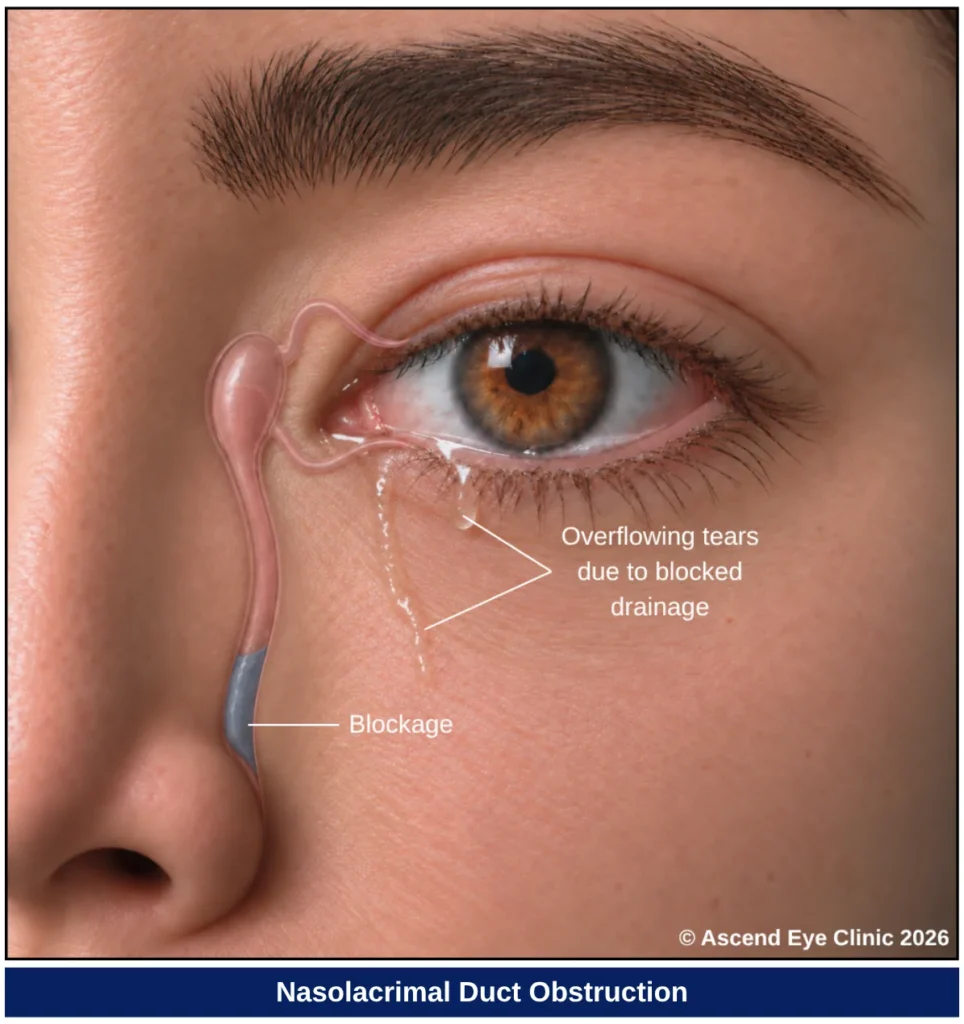
Watery eyes may occur in one or both eyes and can be constant or intermittent. Depending on the underlying cause, they may also occur together with other eye symptoms.
Associated symptoms may include:

Diagnosis begins with a careful assessment of the eyelids, eyelashes and surrounding eye structures. We look at the position and movement of the eyelids, the condition of the lid margins and lashes, and any changes affecting the surface of the eye to determine the nature and extent of the problem.
Measures how clearly you can see and helps identify any reduction in vision.
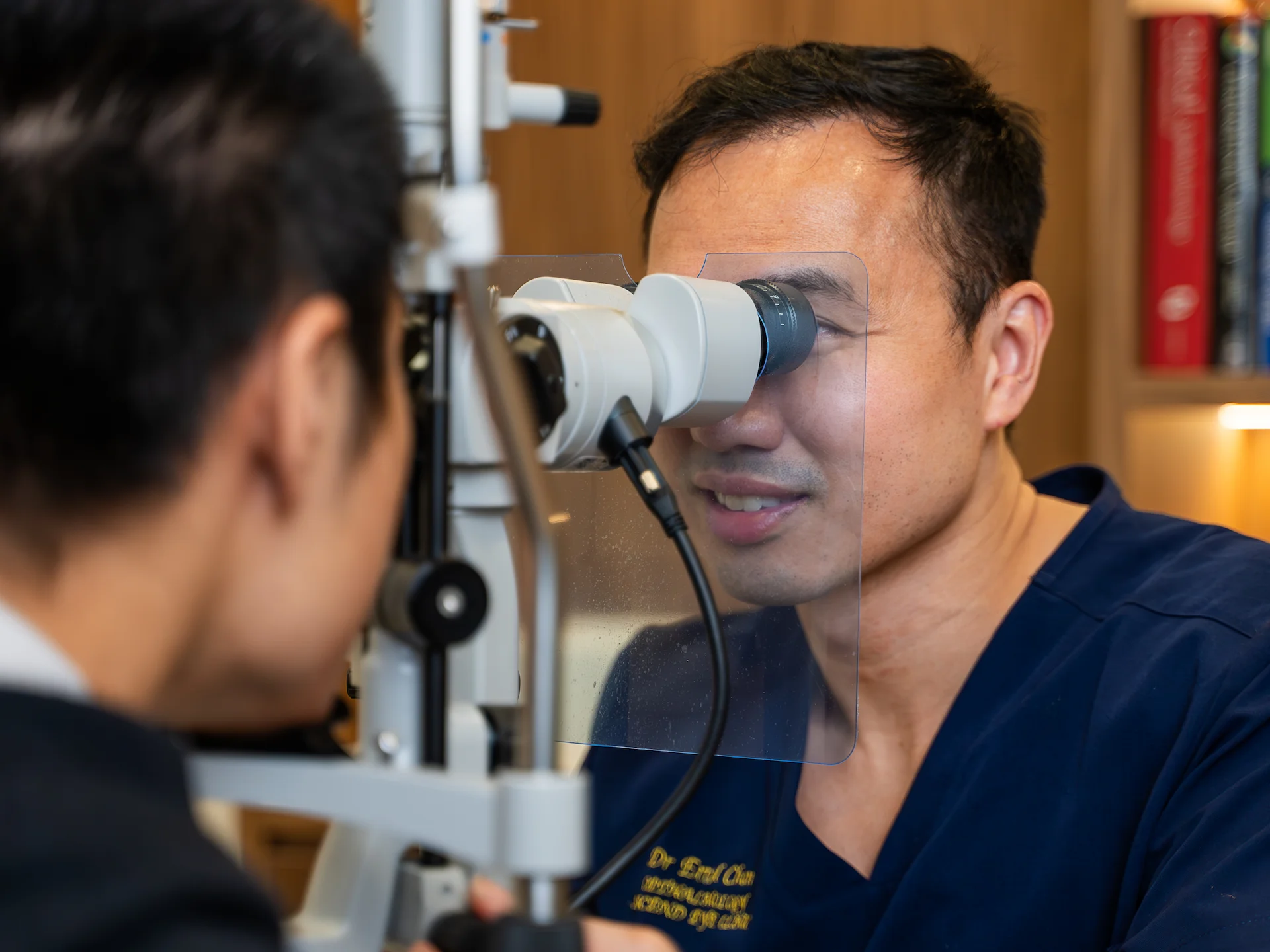
Provides a magnified view of the eyelids, eyelashes, tear film and eye surface to look for irritation, inflammation, eyelid abnormalities or other causes of excessive tearing.
Examines the tear drainage openings and surrounding structures to determine whether excessive tearing is related to narrowing or blockage of the tear drainage system.
Fluid is gently passed through the tear drainage system to assess whether the tear ducts are open or blocked.
Treatment for excessive tearing depends on the underlying cause. We will assess whether the problem arises from the tear drainage system, eyelid position or irritation of the eye surface, then tailor treatment to address the source of the tearing directly.
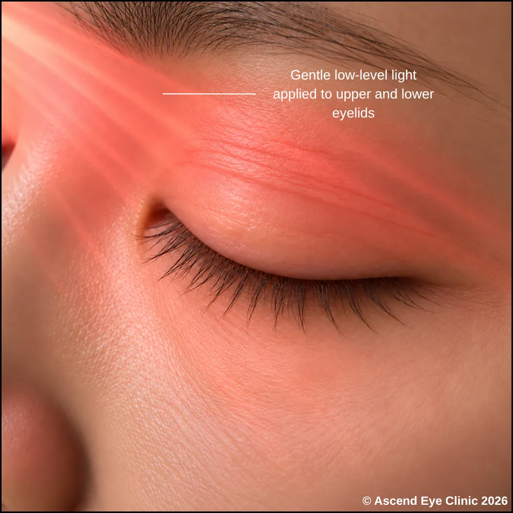
Dry eye treatment aims to stabilise the tear film and reduce the irritation that triggers reflex tearing. Lubricating eye drops replenish moisture on the eye surface, while low-level light therapy (LLLT) supports meibomian gland function and improves the oily layer of the tear film. As the ocular surface becomes more stable and comfortable, the reflex drive for excessive tear production is reduced.
Learn more about our dry eye treatment options here.
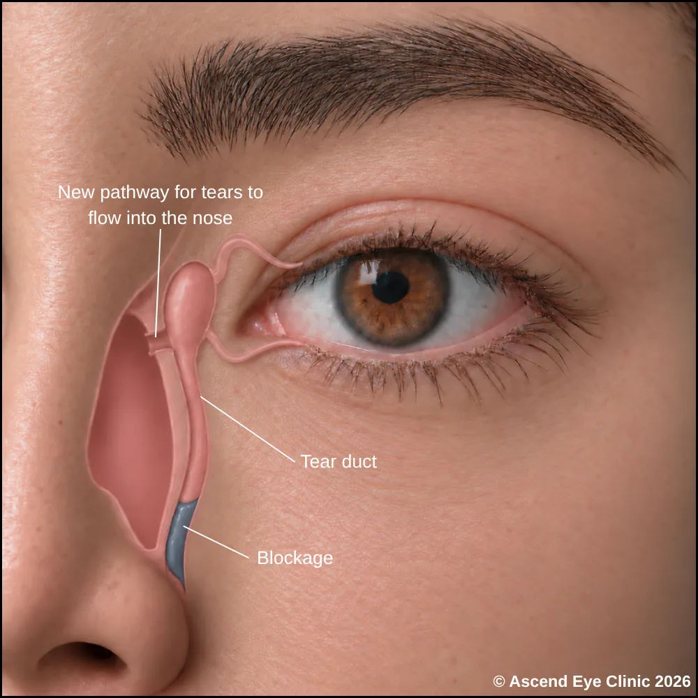
Dacryocystorhinostomy (DCR) is a procedure that creates a new drainage pathway for tears when the nasolacrimal duct is significantly narrowed or blocked. Instead of passing through the blocked duct, tears are redirected from the lacrimal sac directly into the nose through a new opening, allowing them to drain more freely and reducing persistent watering.
The procedure is performed under local anaesthesia with sedation or general anaesthesia. Most DCR procedures are performed endoscopically from inside the nose. A small opening is created between the lacrimal sac and the nasal cavity, and a fine silicone tube may be placed temporarily to support the new passage while it heals.
After the procedure, patients may experience mild swelling, bruising, nasal congestion or a small amount of blood-stained discharge for the first few days. Discomfort is generally mild and can be managed with medication. Follow-up visits are arranged to check that the new drainage passage remains open and to remove the silicone tube if one has been placed.
From your initial consultation to treatment and follow-up care, our team will guide you through every stage of your journey. We will take the time to explain your condition, answer your questions and help you feel informed and supported throughout your care.

Your consultation begins with a detailed assessment of your vision and eye health. Depending on your condition, this may include measurements of your vision and eye pressure, examination of the front and back of the eye, and advanced diagnostic imaging.
Our team will guide you through each test and explain what is being assessed. Your results will then be carefully reviewed to understand your individual needs and determine whether monitoring or treatment may be recommended.
Every eye and every patient is different. Your treatment plan will be tailored according to your examination findings, visual needs, general health and lifestyle.
We will explain the recommended treatment, its expected benefits, possible risks and any suitable alternatives in a clear and straightforward manner. You will have time to ask questions and consider your options before deciding how you would like to proceed.
Before your treatment, our team will guide you through the necessary preparations, paperwork and medication instructions. Where applicable, we will explain how to use your prescribed eye drops and how to care for your eyes afterwards.
We understand that undergoing a procedure can feel unfamiliar or worrying. Our team will be available to answer your questions and ensure that you know what to expect before the day of your treatment.
On the day of your procedure, you will arrive at the hospital or surgical centre where your operation has been scheduled. The surgical team will help you prepare and explain what will happen at each stage.
Anaesthetic eye drops or other forms of anaesthesia may be used, depending on the procedure, to keep you comfortable. Your doctor and the surgical team will support and reassure you throughout the operation.
Most eye surgeries are performed as day procedures, although the duration and recovery process will depend on the treatment you receive. Your vision may be temporarily blurred afterwards, so you should arrange for someone to accompany you home and should not drive unless your doctor has advised that it is safe to do so.
We will provide clear instructions on how to care for your eyes, use any prescribed medication and manage your activities during recovery. We will also explain which symptoms are expected and when you should contact the clinic for further advice.
Follow-up appointments allow us to monitor your vision, assess your recovery and identify any concerns at an early stage. Our team remains accessible should you have any questions or concerns throughout your recovery.
Excessive tearing may be caused by dry eye, irritation or a problem with the tear-drainage system. A detailed examination can help identify the underlying cause and determine the most appropriate treatment.
Book a consultation at Ascend Eye Clinic for an assessment of your eye surface, eyelids and tear-drainage pathway.
Excessive tearing occurs when the eyes produce more tears than needed or when tears cannot drain normally. Causes include dry eye, allergies, eyelid problems, eye surface irritation and narrowing or blockage of the tear drainage system.
When the surface of the eye becomes dry or irritated, the tear glands respond by producing extra watery tears. These reflex tears do not provide the same stable lubrication as a healthy tear film and can overflow onto the cheeks.
Persistent watering in one eye can occur when there is a problem affecting that eye or its tear drainage pathway. A narrowed or blocked tear duct, eyelid position problem, misdirected eyelash or local eye surface irritation are possible causes.
Wind, dry air, air-conditioning, smoke and other environmental triggers can irritate the eye surface and stimulate reflex tearing. This is particularly noticeable when the tear film is unstable or the eyes are dry.
Dry eye often causes grittiness, burning, fluctuating vision or sensitivity to wind and air-conditioning. A blocked tear duct tends to cause more persistent overflowing of tears and can affect one eye more than the other. An examination helps determine the actual cause.
Yes. When tears cannot drain properly, fluid and mucus can collect within the tear drainage system. This can lead to sticky discharge around the inner corner of the eye.
Yes. Stagnant tears within the lacrimal sac can allow bacteria to multiply and cause dacryocystitis. This causes redness, pain, tenderness and swelling near the inner corner of the eye and requires medical treatment.
The eyelids, puncta and surface of the eye are examined first. The tear drainage pathway can then be gently irrigated with fluid to assess whether it passes freely into the nose and to identify narrowing or blockage.
Eye drops can reduce tearing when the cause is dry eye, allergy or inflammation of the eye surface. They do not reopen a significantly blocked nasolacrimal duct, so treatment depends on the underlying cause.
Treatment depends on where the narrowing occurs and how severe it is. A narrowed punctum can be enlarged, and some blockages can be managed with probing or silicone tube placement. A significant blockage of the nasolacrimal duct can require dacryocystorhinostomy, or DCR.
Dacryocystorhinostomy (DCR) creates a new drainage passage between the lacrimal sac and the inside of the nose. This bypasses the blocked nasolacrimal duct so tears have another route to drain.
DCR can be performed through a small incision beside the nose or endoscopically from inside the nose. A new opening is created between the lacrimal sac and the nasal cavity, and a fine silicone tube can be temporarily placed to support the new drainage passage while it heals.
DCR is performed under local anaesthesia with sedation or under general anaesthesia. The type of anaesthesia depends on the surgical approach and individual requirements.
Mild swelling, bruising, nasal congestion and some blood-stained tears or nasal discharge can occur during the early recovery period. Some watering can also continue initially while swelling settles and the new drainage passage heals.
Assessment is recommended when watering is persistent, repeatedly runs down the cheek, mainly affects one eye, interferes with vision or daily activities, or occurs together with discharge or swelling. Painful redness and swelling near the inner corner of the eye should be assessed promptly.
Hi, I would like to book a consultation with Dr Errol Chan from Ascend Eye Clinic
