
A sudden increase in floaters or flashes can be an early warning sign of a retinal tear or detachment and should not be ignored. Prompt retinal assessment allows us to identify the cause quickly and treat any retinal problem before more serious vision loss develops.


Floaters most commonly develop as the vitreous, the clear gel inside the eye, naturally changes with age and begins to separate from the retina. This process is known as a posterior vitreous detachment (PVD). Floaters can also occur with eye trauma, inflammation, and various retinal conditions.
Flashes are usually caused by the vitreous pulling on the retina during this separation. In most cases, the traction settles without causing damage. However, if the pulling is strong enough, it can create a retinal tear, which can progress to a retinal detachment if not treated promptly.
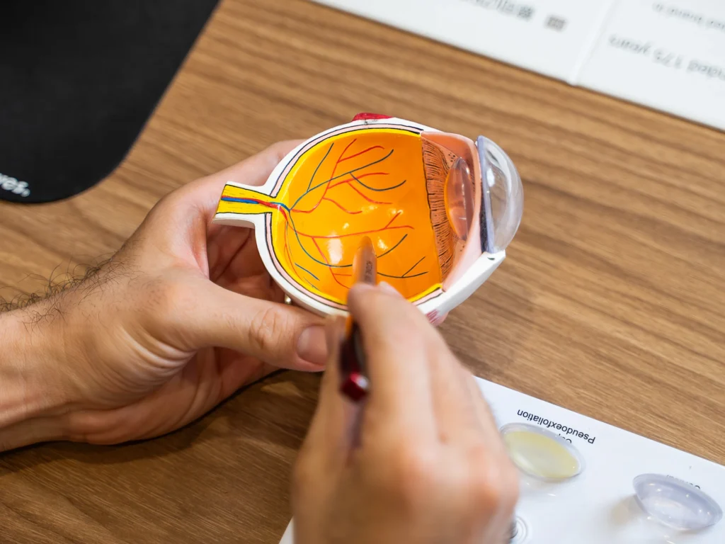
Many longstanding floaters are harmless. However, seek an urgent eye assessment if you experience:
These symptoms may indicate a retinal tear or retinal detachment, which requires prompt treatment to protect your vision.
We use a combination of detailed eye examination and advanced retinal imaging to assess the macula, retina, optic nerve, and retinal blood vessels. These tests allow us to identify structural and functional changes, determine the type and severity of the condition, and establish a clear baseline for ongoing monitoring and treatment planning.
Measures how clearly you can see and helps identify any reduction in central vision.
Examines the vitreous for floaters, opacities and signs of vitreous detachment, while assessing the retina for associated abnormalities.
Captures detailed images of the retina, including the peripheral retina, to identify retinal tears, retinal detachment, vitreous haemorrhage and other associated abnormalities.
Provides detailed cross-sectional images of the retina to assess whether the vitreous remains attached to the retina or has separated, particularly around the macula.
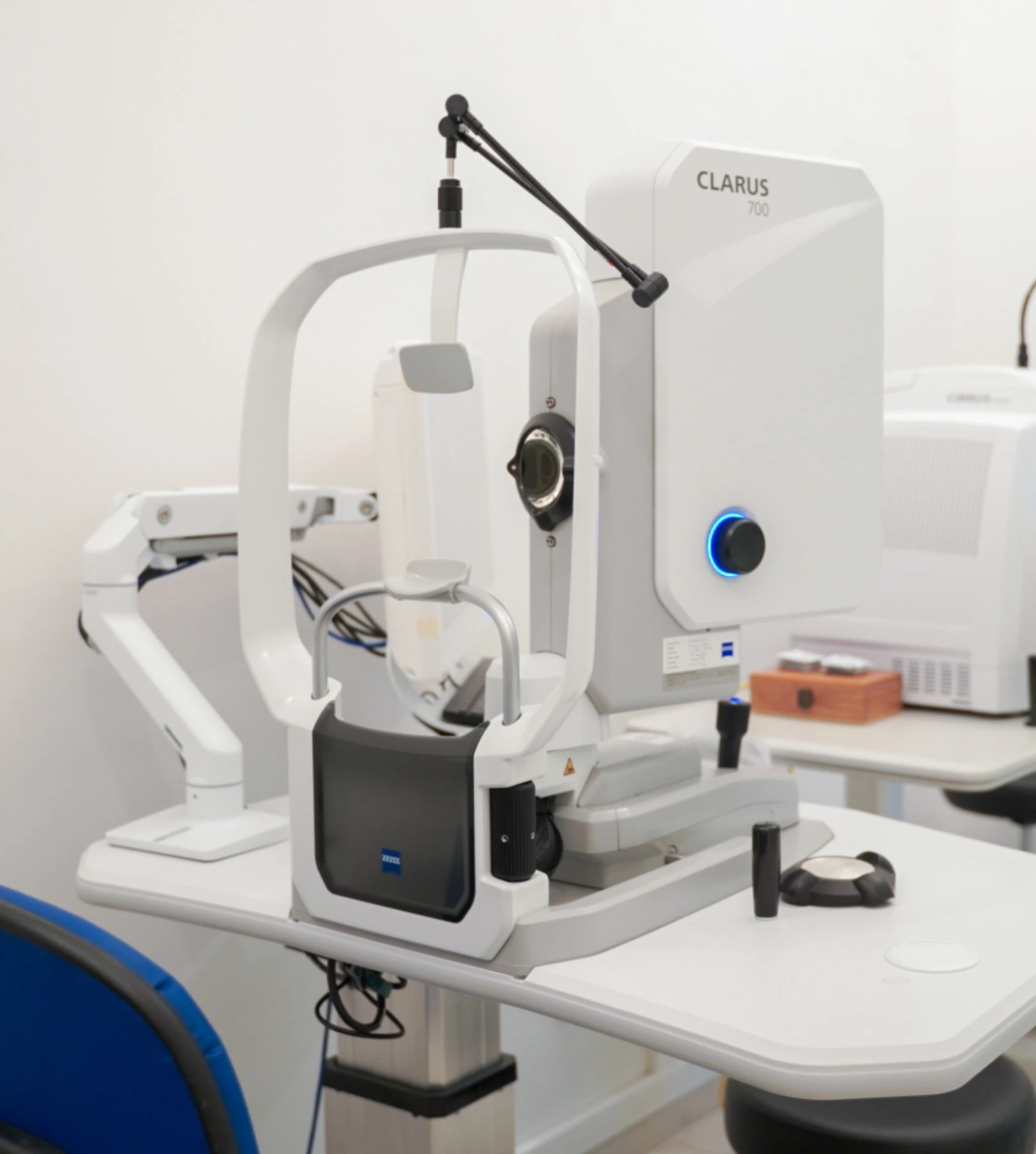
Many floaters and flashes do not require treatment, particularly when they are caused by an uncomplicated posterior vitreous detachment. However, if we identify a retinal tear, retinal detachment, or another underlying retinal problem, treatment should be performed promptly.
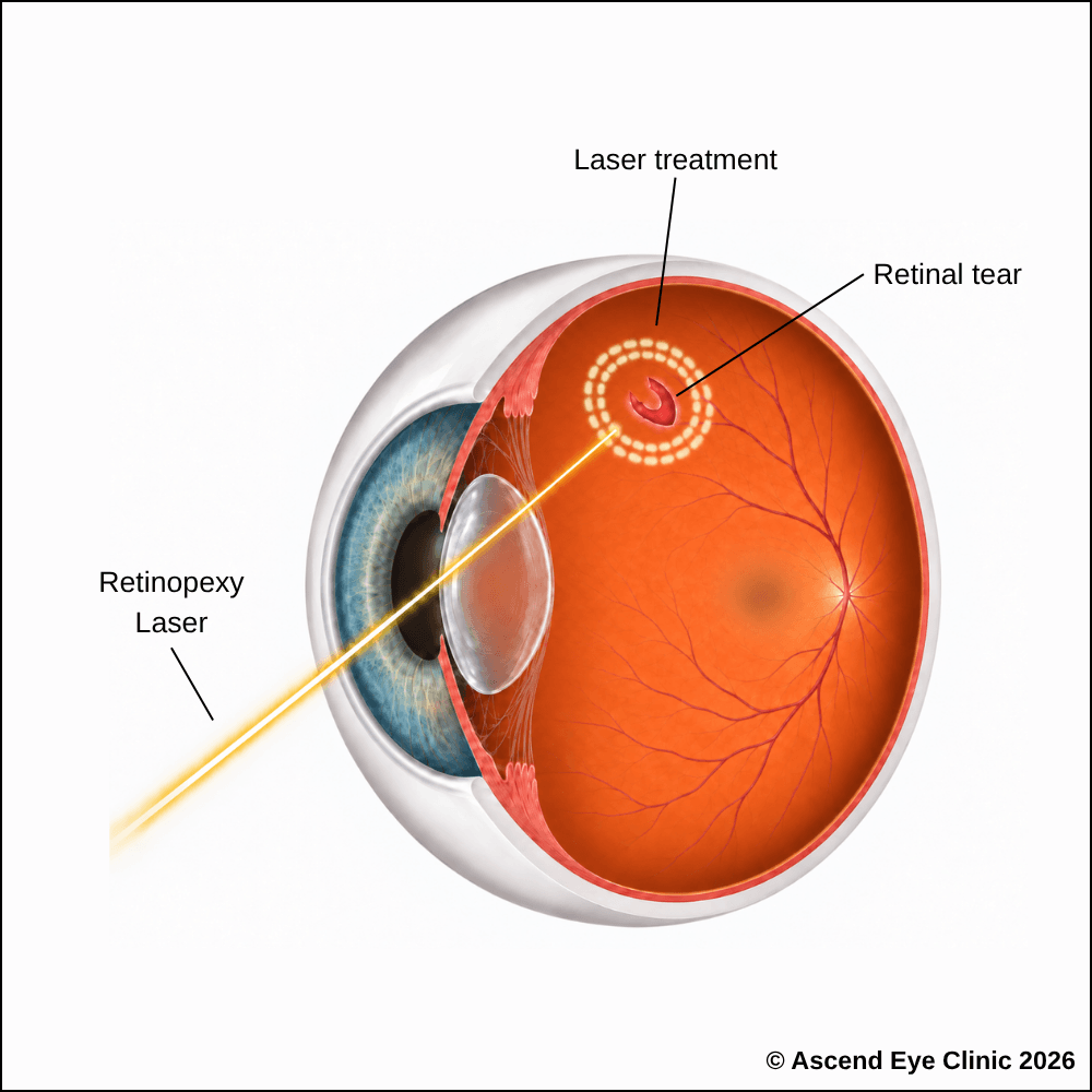
When the vitreous pulls on the retina, it can cause a retinal tear, resulting in the sudden appearance of floaters and flashes. Retinal tears require prompt treatment to prevent progression to retinal detachment.
Laser retinopexy is used to secure the retina around a retinal tear or hole and reduce the risk of it progressing into a retinal detachment. The laser creates a series of small, controlled treatment spots around the affected area, which develop into a firm adhesion between the retina and the underlying tissue. This forms a protective barrier that helps prevent fluid from passing through the tear and lifting the retina further.
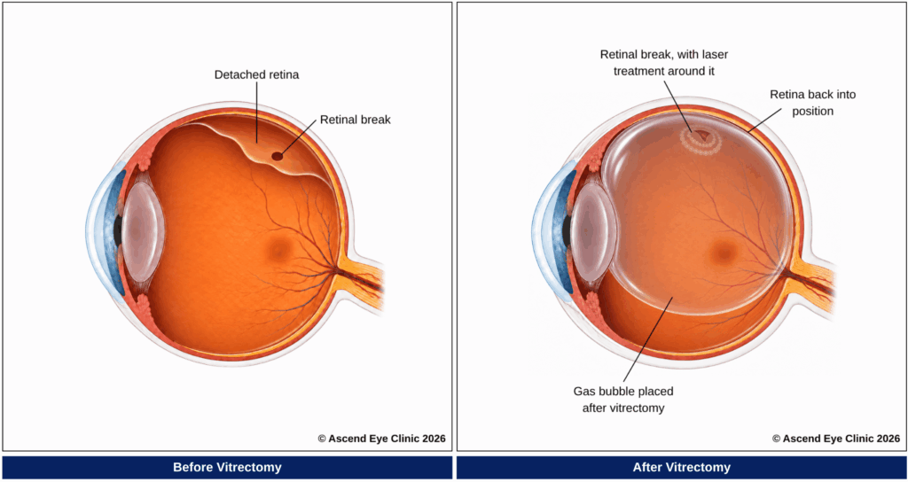
If a retinal tear progresses to retinal detachment, surgical treatment is required to reattach the retina and preserve vision. Depending on the extent and location of the detachment, treatment includes pneumatic retinopexy, scleral buckling or vitrectomy.
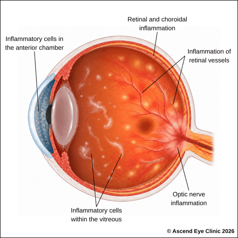
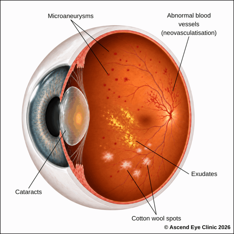
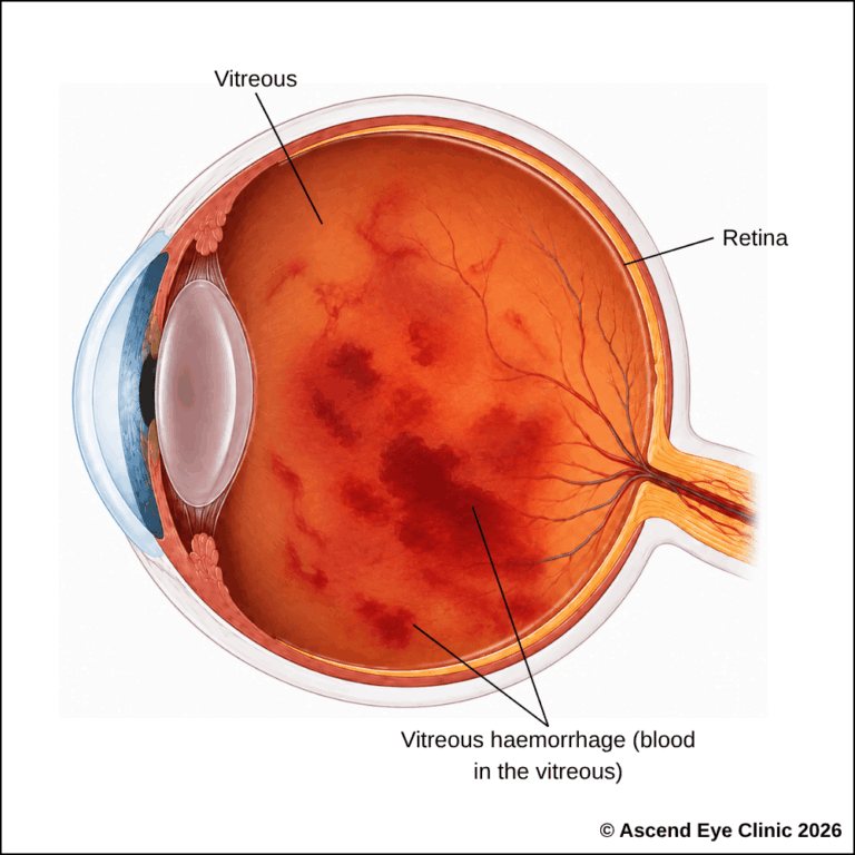
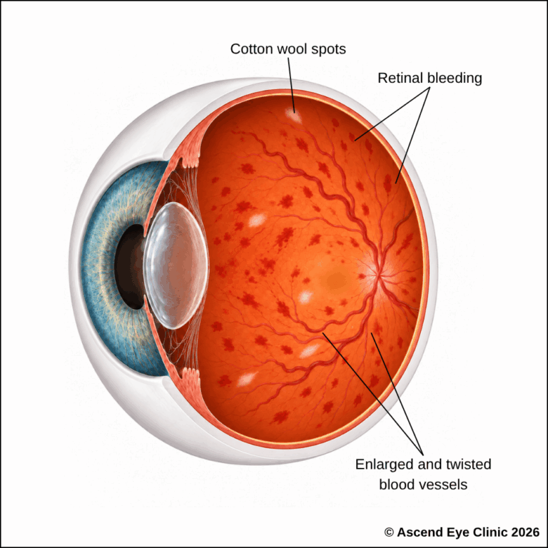
Floaters can also be caused by bleeding inside the eye or inflammation, including in conditions such as:
In these cases, treatment focuses on controlling the underlying condition and clearing the cause of the floaters. This can include medication, retinal laser, injections or vitrectomy, depending on what is found during the examination.
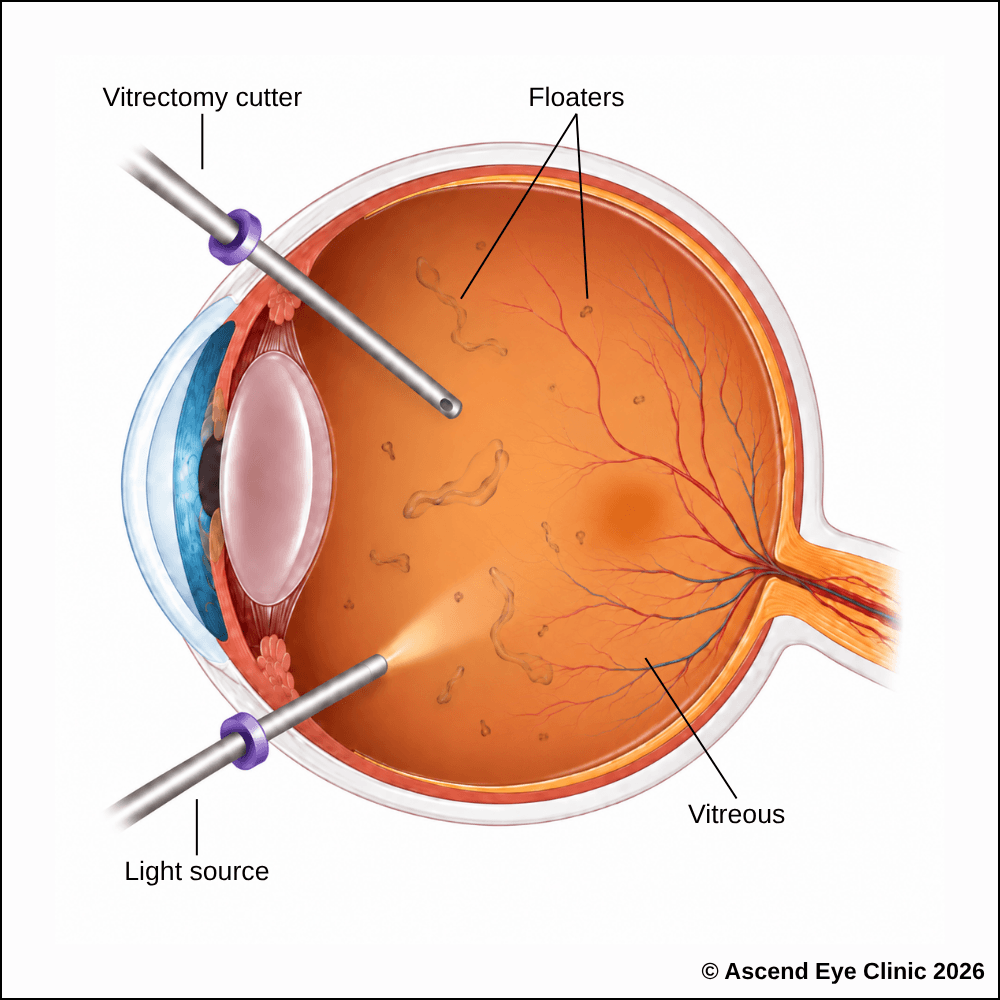
For severe floaters that remain visually disruptive and interfere with activities such as reading, driving or working on screens, vitrectomy can be performed to remove the vitreous gel containing the floaters from inside the eye. By removing the material casting shadows across the retina, the procedure aims to provide a clearer and less obstructed field of vision.
Vitrectomy is performed in an operating theatre through very small openings in the eye using fine microsurgical instruments. The vitreous gel is carefully removed while a clear fluid is used to maintain the shape and pressure of the eye during surgery. Once the vitreous and troublesome floaters have been cleared, the eye is left filled with clear fluid, which is gradually replaced by the eye’s own natural fluid.
Anaesthesia is used to keep the eye comfortable throughout the procedure. Vision is blurred initially and gradually becomes clearer as the eye recovers, with follow-up visits used to monitor healing and the retina.
From your initial consultation to treatment and follow-up care, our team will guide you through every stage of your journey. We will take the time to explain your condition, answer your questions and help you feel informed and supported throughout your care.

Your consultation begins with a detailed assessment of your vision and eye health. Depending on your condition, this may include measurements of your vision and eye pressure, examination of the front and back of the eye, and advanced diagnostic imaging.
Our team will guide you through each test and explain what is being assessed. Your results will then be carefully reviewed to understand your individual needs and determine whether monitoring or treatment may be recommended.
Every eye and every patient is different. Your treatment plan will be tailored according to your examination findings, visual needs, general health and lifestyle.
We will explain the recommended treatment, its expected benefits, possible risks and any suitable alternatives in a clear and straightforward manner. You will have time to ask questions and consider your options before deciding how you would like to proceed.
Before your treatment, our team will guide you through the necessary preparations, paperwork and medication instructions. Where applicable, we will explain how to use your prescribed eye drops and how to care for your eyes afterwards.
We understand that undergoing a procedure can feel unfamiliar or worrying. Our team will be available to answer your questions and ensure that you know what to expect before the day of your treatment.
On the day of your procedure, you will arrive at the hospital or surgical centre where your operation has been scheduled. The surgical team will help you prepare and explain what will happen at each stage.
Anaesthetic eye drops or other forms of anaesthesia may be used, depending on the procedure, to keep you comfortable. Your doctor and the surgical team will support and reassure you throughout the operation.
Most eye surgeries are performed as day procedures, although the duration and recovery process will depend on the treatment you receive. Your vision may be temporarily blurred afterwards, so you should arrange for someone to accompany you home and should not drive unless your doctor has advised that it is safe to do so.
We will provide clear instructions on how to care for your eyes, use any prescribed medication and manage your activities during recovery. We will also explain which symptoms are expected and when you should contact the clinic for further advice.
Follow-up appointments allow us to monitor your vision, assess your recovery and identify any concerns at an early stage. Our team remains accessible should you have any questions or concerns throughout your recovery.
New floaters and flashes are often caused by changes within the vitreous, but they can also be an early sign of a retinal tear or retinal detachment.
Book a consultation at Ascend Eye Clinic for a comprehensive dilated retinal examination. We will identify the likely cause of your symptoms and advise whether monitoring or treatment is required.
Floaters develop when small strands or clumps form within the vitreous, the clear gel that fills the eye. As light passes through the eye, these structures cast shadows onto the retina, which are seen as spots, threads, cobwebs or drifting shapes.
A sudden increase in floaters commonly occurs when the vitreous changes with age and separates from the retina, known as a posterior vitreous detachment. New floaters should be examined promptly because vitreous separation can occasionally pull strongly enough to create a retinal tear.
Yes. Floaters become increasingly common as the vitreous naturally becomes more liquid and develops small condensations with age. However, a sudden increase in floaters is different from long-standing stable floaters and should be assessed.
Floaters can appear as small dots, strands, rings, cobwebs or translucent shapes that drift across the vision. They are often easiest to notice against bright, plain backgrounds such as a white wall, computer screen or clear sky.
Floaters are suspended within the vitreous, so they move as the vitreous shifts when the eyes move. They often continue drifting briefly after the eye stops moving, which gives them their characteristic floating appearance.
Many floaters become much less noticeable over time. The floaters themselves do not necessarily disappear completely, but they can move away from the central visual axis and the brain gradually becomes less aware of them.
Long-standing floaters often remain stable or become less noticeable. A sudden increase in number, especially if accompanied by flashes, blurred vision or a shadow in the vision, requires prompt examination to exclude a retinal tear or detachment.
Flashes occur when the vitreous pulls or tugs on the retina, mechanically stimulating the retinal cells and creating the sensation of light even though no external light is present. This commonly happens during a posterior vitreous detachment.
Flashes are easier to notice in dim surroundings because there is less background light competing with them. They often appear as brief streaks, arcs or flickers at the side of the vision.
No. Posterior vitreous detachment is a common cause, but floaters and flashes can also occur with retinal tears, retinal detachment, bleeding inside the eye, inflammation, trauma and other retinal conditions. A detailed retinal examination identifies the underlying cause.
Sudden new floaters, a marked increase in existing floaters, repeated flashes, blurred vision or any new shadow or missing area of vision should be assessed urgently. These symptoms can occur when the vitreous is pulling strongly on the retina or when a retinal tear has developed.
A sudden shower of numerous small floaters can indicate pigment cells or blood entering the vitreous after a retinal tear. This is an important warning symptom and requires an urgent dilated retinal examination.
Seek urgent eye assessment immediately. A new curtain, shadow or expanding dark area can indicate that the retina has detached, and early treatment is important to protect as much vision as possible.
The pattern of symptoms provides useful clues, but the only reliable way to determine the cause is through a detailed examination of the retina. We pay particular attention to sudden onset, increasing symptoms, flashes, reduced vision, vitreous bleeding and any shadow or field loss.
Sudden new floaters or flashes should be assessed promptly, particularly if the symptoms are increasing or affecting only one eye. A dilated retinal examination allows us to check the peripheral retina carefully for tears, bleeding or early retinal detachment.
Yes, particularly with a high minus prescription. Highly myopic eyes are usually longer and have a thinner, more stretched retina, which increases the likelihood of earlier vitreous separation, peripheral retinal degeneration, retinal tears and retinal detachment.
Yes. Changes within the eye after surgery can make pre-existing floaters more noticeable or contribute to vitreous separation. Any sudden onset of new floaters, flashes or a shadow after eye surgery should be examined promptly.
Yes. Trauma can cause the vitreous to pull on the retina and can lead to retinal tears, bleeding or retinal detachment. New floaters or flashes following an eye injury warrant a careful retinal examination even if the vision initially seems normal.
If the retina is healthy and no tear, bleeding or inflammation is present, the floaters are usually caused by harmless changes within the vitreous itself. They can remain visible for months or longer, although most people notice them progressively less as they settle and the brain adapts.
Most floaters do not require treatment because they gradually become less intrusive. When dense, persistent floaters significantly interfere with reading, driving or daily activities, treatment can be considered after a detailed assessment; vitrectomy is the most definitive option because it removes the vitreous gel containing the floaters.
Hi, I would like to book a consultation with Dr Errol Chan from Ascend Eye Clinic
