
A retinal vein occlusion occurs when there is a sudden obstruction of blood flow in the retinal veins. A retinal vein occlusion suddenly disrupts blood flow within the retina and leads to visual loss. A retinal vein occlusion can also cause longer term complications such as retinal swelling or chronic lack of blood flow to the retina. Prompt assessment and appropriate management are important to limit retinal damage, manage associated complications, and optimise visual outcomes.
A retinal vein occlusion occurs when a vein responsible for carrying blood away from the retina becomes blocked. This causes pressure to build within the retinal circulation, leading to bleeding and leakage of fluid into the surrounding tissue.
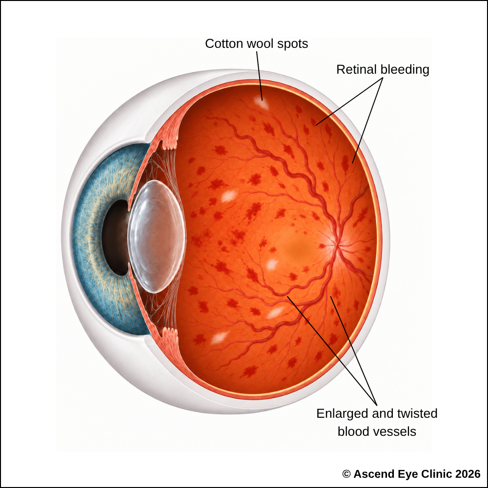
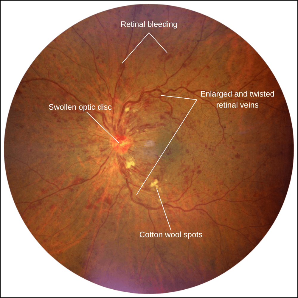
A Central Retinal Vein Occlusion occurs when the main vein draining blood from the retina becomes blocked. Because this vein drains the entire retina, retinal haemorrhages, vessel congestion, and swelling can be more widespread than in BRVO.
CRVO can cause significant reduction in vision, particularly when macular oedema develops or when retinal circulation is severely reduced.
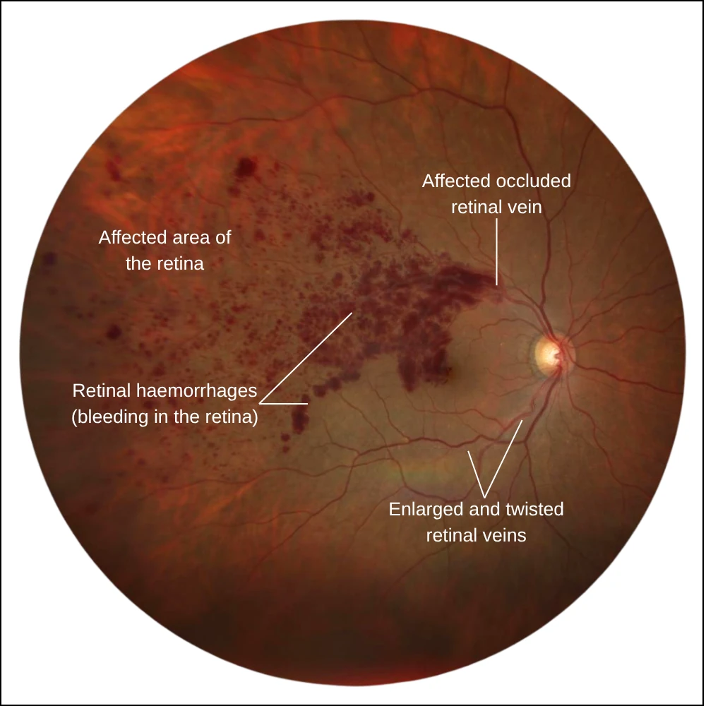
A Branch Retinal Vein Occlusion occurs when one of the smaller veins draining a section of the retina becomes blocked. As a result, retinal haemorrhages and swelling are usually confined to the area supplied by the affected vein.
Vision is particularly affected when the blockage causes fluid to leak into the macula, resulting in macular oedema. The level of visual disturbance depends on the location of the affected vein, the amount of macular swelling and whether part of the retina has received insufficient blood flow.
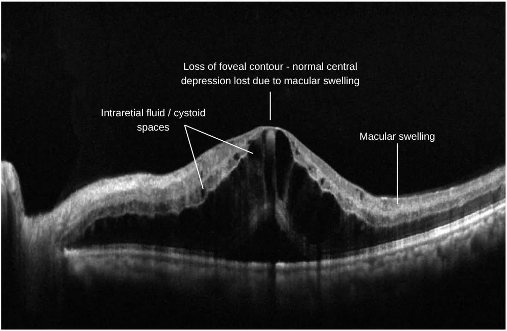
Macular oedema is one of the most important causes of reduced vision in retinal vein occlusion. When a retinal vein becomes blocked, pressure builds within the affected blood vessels and disrupts their normal ability to contain fluid. Fluid then leaks into the surrounding retina and can accumulate within the macula, causing it to become swollen.
Because the macula is responsible for sharp central vision, this swelling can cause blurred or distorted vision, reduced contrast, and difficulty recognising faces.
Retinal vein occlusions are more common in people with conditions that affect the health of the blood vessels and circulation. These conditions can increase the likelihood of reduced blood flow or blockage within the retinal veins.
Common risk factors include:

A retinal vein occlusion can cause retinal swelling, bleeding and reduced blood flow. Symptoms vary depending on which retinal vein is affected and may include:



If the macula is affected by swelling, central vision may become blurred or distorted. In branch retinal vein occlusion, the visual disturbance may affect only part of the visual field, depending on the location of the blocked vein.
We use a combination of detailed eye examination and advanced retinal imaging to assess the macula, retina, optic nerve, and retinal blood vessels. These tests allow us to identify structural and functional changes, determine the type and severity of the condition, and establish a clear baseline for ongoing monitoring and treatment planning.
Measures how clearly you can see and helps identify any reduction in central vision.
Captures detailed images of the retina and retinal blood vessels to document retinal haemorrhages, dilated or tortuous veins and other changes caused by the occlusion.
Uses fluorescein dye and a series of retinal photographs to assess retinal blood flow, identify areas of poor circulation or non-perfusion, and detect leakage or abnormal new blood vessels.
Provides cross-sectional images of the retina to assess the macula for swelling, intraretinal fluid and other structural changes associated with retinal vein occlusion.
Provides detailed images of the retinal blood vessels without dye to assess blood flow, identify areas of reduced perfusion and evaluate changes in the retinal capillary network.
Maps the central and peripheral visual field to identify areas of reduced or missing vision associated with the affected part of the retina.
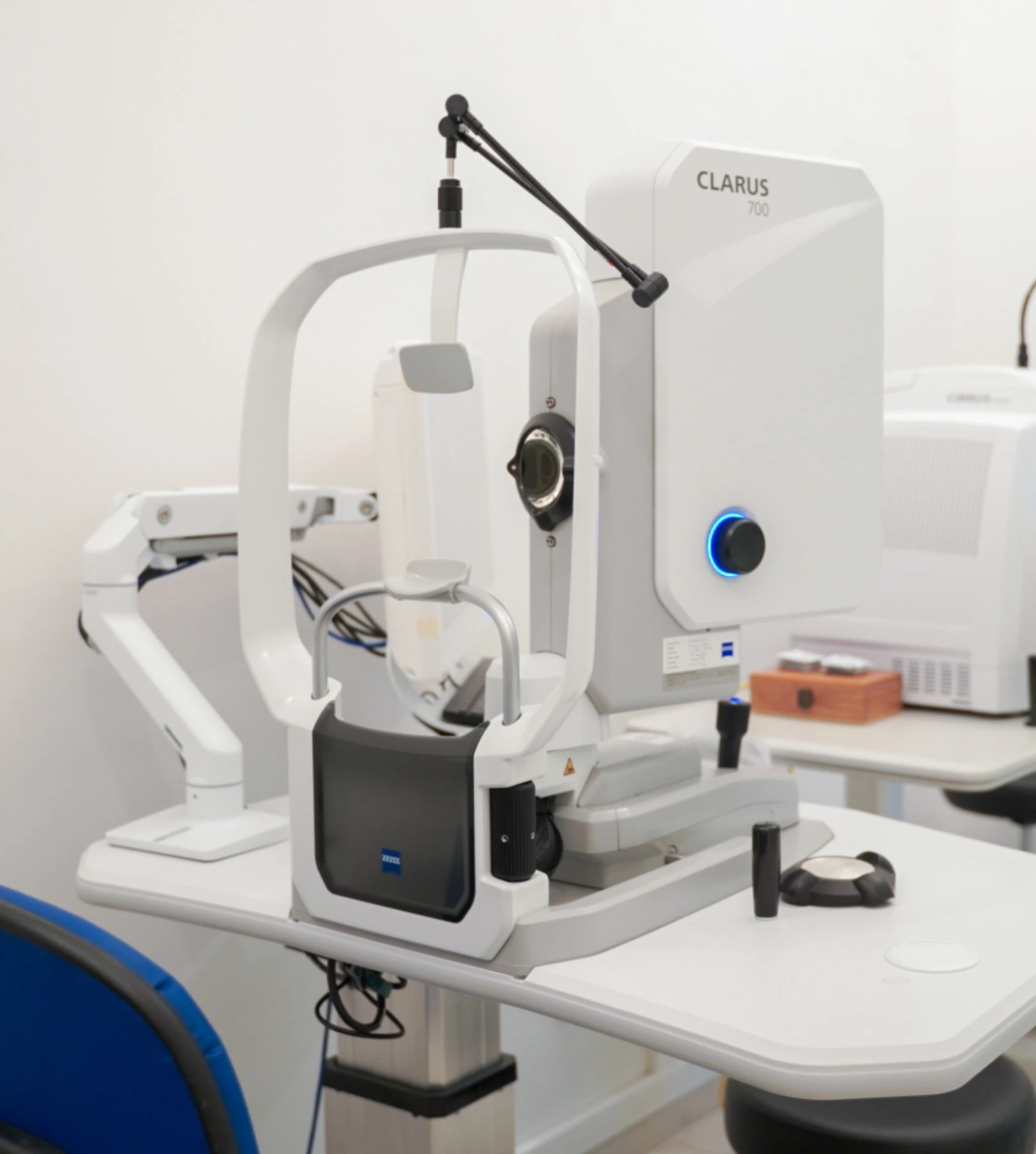
Treatment focuses on reducing macular oedema, protecting vision, and lowering the risk of further retinal damage. The most appropriate treatment is based on where the blockage has occurred, how much of the retina is affected by reduced blood flow, and the severity of the macular swelling.
Anti-VEGF injections are the main treatment for macular oedema caused by retinal vein occlusion. The anti-VEGF medication is injected into the eye to reduce fluid leakage and swelling in the macula, helping to improve or stabilise vision.
At Ascend Eye Clinic, anti-VEGF medications available include Eylea®, Lucentis®, and Vabysmo®. We will advise on the most appropriate medication based on your condition and treatment needs.
An intravitreal injection is performed as a quick outpatient procedure with careful preparation to keep the eye comfortable throughout. Anaesthetic drops are used to thoroughly numb the eye before the injection. Although the idea of an injection into the eye can sound daunting, the injection itself takes only a few seconds, much like a vaccination procedure, and most people feel only very mild discomfort.
Several injections are often given at regular intervals over several months to bring the retinal condition under control and achieve stability. Active treatment can be stopped and maintainence treatment can be followed with continually monitoring the retina closely with imaging during follow-up visits.
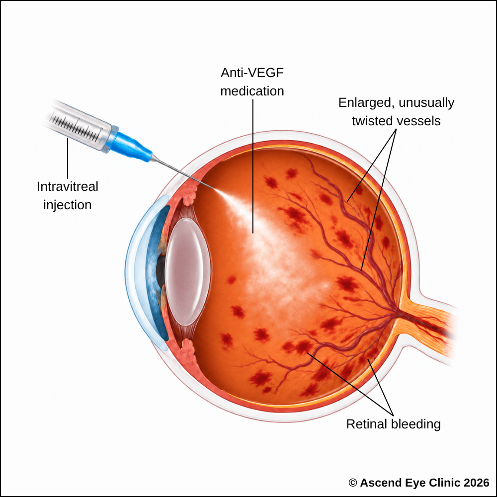
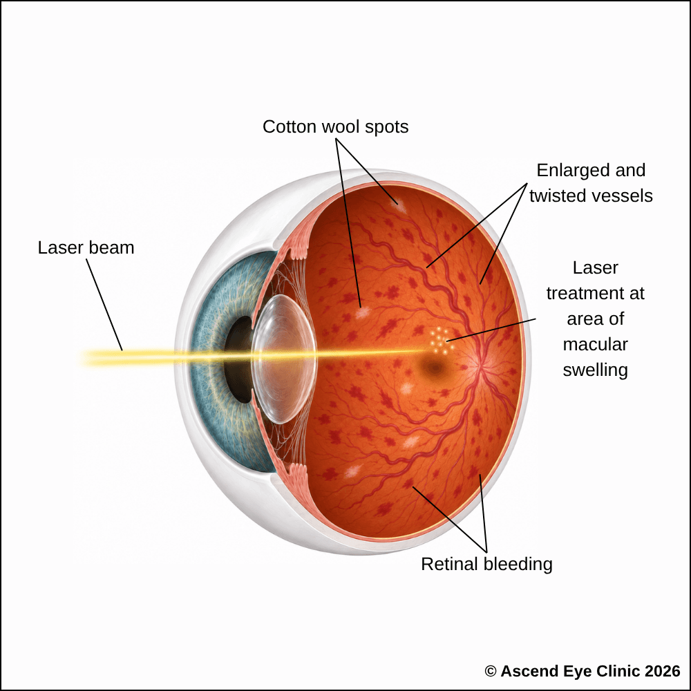
Macular laser is a treatment used in selected cases of macular oedema caused by retinal vein occlusion, where damaged retinal blood vessels are leaking fluid into the macula. The laser applies small, controlled spots to these leaking areas while avoiding the centre of the macula responsible for detailed vision.
Treating these areas helps reduce further fluid leakage and stabilise swelling within the macula. Macular laser laser is used when swelling persists or when the pattern of leakage is suitable for targeted laser treatment.
Macular laser treatment is performed in a laser treatment room with anaesthetic drops used to numb the eye and keep you comfortable throughout the procedure. A special contact lens is then gently placed on the surface to guide the laser accurately. You will see repeated flashes of light as multiple laser spots are applied across the macular area. Treatment is generally well tolerated, although some people may experience mild discomfort during the procedure.
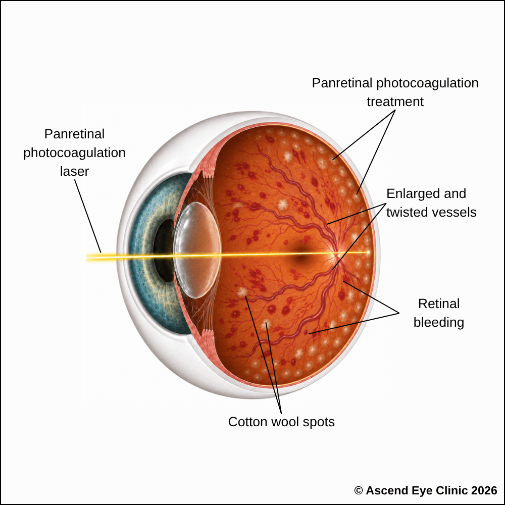
Panretinal photocoagulation (PRP) is a laser treatment used when parts of the retina are not receiving enough blood and oxygen because the retinal circulation has been damaged or obstructed. The laser creates many small, controlled spots in these unhealthy areas of the peripheral retina, while leaving the central retina responsible for detailed vision untreated.
Treating these oxygen-deprived areas reduces the retina’s production of vascular endothelial growth factor, or VEGF, a protein that stimulates the growth of fragile and abnormal blood vessels. By lowering VEGF levels, PRP reduces the likelihood of further abnormal blood vessel growth and bleeding inside the eye, while helping prevent complications such as neovascular glaucoma, which can cause a dangerous rise in eye pressure.
PRP laser treatment is performed in a laser treatment room with anaesthetic drops used to numb the eye and keep you comfortable throughout the procedure. A special contact lens is then gently placed on the surface to guide the laser accurately. You will see repeated flashes of light as multiple laser spots are applied across the peripheral retina. Treatment is generally well tolerated, although some people may experience mild discomfort during the procedure.
From your initial consultation to treatment and follow-up care, our team will guide you through every stage of your journey. We will take the time to explain your condition, answer your questions and help you feel informed and supported throughout your care.

Your consultation begins with a detailed assessment of your vision and eye health. Depending on your condition, this may include measurements of your vision and eye pressure, examination of the front and back of the eye, and advanced diagnostic imaging.
Our team will guide you through each test and explain what is being assessed. Your results will then be carefully reviewed to understand your individual needs and determine whether monitoring or treatment may be recommended.
Every eye and every patient is different. Your treatment plan will be tailored according to your examination findings, visual needs, general health and lifestyle.
We will explain the recommended treatment, its expected benefits, possible risks and any suitable alternatives in a clear and straightforward manner. You will have time to ask questions and consider your options before deciding how you would like to proceed.
Before your treatment, our team will guide you through the necessary preparations, paperwork and medication instructions. Where applicable, we will explain how to use your prescribed eye drops and how to care for your eyes afterwards.
We understand that undergoing a procedure can feel unfamiliar or worrying. Our team will be available to answer your questions and ensure that you know what to expect before the day of your treatment.
On the day of your procedure, you will arrive at the hospital or surgical centre where your operation has been scheduled. The surgical team will help you prepare and explain what will happen at each stage.
Anaesthetic eye drops or other forms of anaesthesia may be used, depending on the procedure, to keep you comfortable. Your doctor and the surgical team will support and reassure you throughout the operation.
Most eye surgeries are performed as day procedures, although the duration and recovery process will depend on the treatment you receive. Your vision may be temporarily blurred afterwards, so you should arrange for someone to accompany you home and should not drive unless your doctor has advised that it is safe to do so.
We will provide clear instructions on how to care for your eyes, use any prescribed medication and manage your activities during recovery. We will also explain which symptoms are expected and when you should contact the clinic for further advice.
Follow-up appointments allow us to monitor your vision, assess your recovery and identify any concerns at an early stage. Our team remains accessible should you have any questions or concerns throughout your recovery.
If your vision suddenly becomes blurred, distorted, or partly missing, seek an eye assessment promptly. Early diagnosis of a retinal vein or artery occlusion allows treatment and monitoring to begin as soon as possible, helping to protect your remaining vision.
A retinal vein occlusion requires prompt assessment, particularly when there is a sudden reduction or marked blurring of vision. Early examination allows us to confirm the diagnosis, assess whether the macula is swollen and identify areas of reduced retinal circulation or other complications that require treatment or close monitoring.
Retinal vein occlusions are typically painless. Most patients notice a sudden change in vision without redness, discomfort or other warning symptoms. Any sudden unexplained reduction or deterioration in vision should therefore be assessed promptly even when the eye feels completely comfortable.
A branch retinal vein occlusion occurs when one of the smaller veins draining a particular area of the retina becomes blocked. The visual effect therefore depends on which part of the retina is involved. A central retinal vein occlusion affects the main retinal vein and involves a much larger area of the retina, often causing more extensive retinal haemorrhage, swelling and visual disturbance.
Symptoms include sudden blurred or reduced vision, distortion, a dark or missing area within the field of vision, or a general reduction in visual clarity. The extent of visual loss depends on the location and severity of the blockage and whether the macula, which is responsible for detailed central vision, is affected.
Yes. Permanent visual loss can occur when the retina has sustained significant damage or when complications such as persistent macular oedema or retinal ischaemia develop. The visual outcome varies according to the type and severity of the vein occlusion, which is why appropriate treatment and follow-up are important.
Yes. A branch retinal vein occlusion can affect only the portion of retina drained by the blocked vein. This can produce blurring, distortion or a missing area in one part of the visual field while the remainder of the vision remains relatively unaffected.
A retinal vein occlusion occurs when blood flow through a retinal vein becomes obstructed. This is often related to changes in the retinal blood vessels associated with ageing, high blood pressure, diabetes, high cholesterol and other vascular conditions. In some cases, compression of a retinal vein by a neighbouring artery also contributes to the blockage.
Risk increases with age and is higher in people with high blood pressure, diabetes, high cholesterol and cardiovascular disease. Smoking, glaucoma, raised eye pressure and certain blood or clotting disorders are also associated with an increased risk of retinal vein occlusion.
Yes. High blood pressure, diabetes and high cholesterol are important risk factors for retinal vein occlusion. These conditions affect blood vessels throughout the body, including the delicate circulation of the retina. We therefore consider both the retinal findings and the patient's general vascular health when assessing the condition.
Yes. Another retinal vein occlusion can develop in the same eye or in the other eye, particularly when underlying vascular risk factors remain poorly controlled. Managing blood pressure, blood sugar, cholesterol and other relevant medical conditions is an important part of reducing future vascular risk.
Yes. Although many patients experience a retinal vein occlusion in only one eye, the underlying vascular risk factors affect the circulation throughout the body and can therefore place the other eye at risk. Continued management of these risk factors remains important even after the affected eye has stabilised.
Visual recovery depends on the type and severity of the occlusion and how extensively the macula has been affected. Vision often improves as retinal haemorrhage and swelling resolve, particularly when macular oedema responds well to treatment. More severe retinal ischaemia or permanent damage to the macula can limit the amount of visual recovery.
Treatment addresses complications of retinal vein occlusion, particularly macular oedema and abnormal new blood vessel growth. However, the original blockage can leave lasting changes within the retina. Vision can therefore remain reduced even when the condition has stabilised. We monitor the retinal structure closely to determine whether there is ongoing swelling or whether the remaining visual limitation reflects permanent retinal damage.
Eye injections are commonly used when a retinal vein occlusion causes macular oedema and reduces central vision. Anti-VEGF medication is injected into the eye to reduce vascular leakage and swelling within the macula. A series of injections is often required initially, with the interval adjusted according to the response seen on examination and OCT imaging.
We assess the vision and examine the retina carefully for the characteristic pattern of retinal haemorrhage, dilated or tortuous veins, swelling and other vascular changes. OCT imaging allows us to assess the macula in detail, while retinal photography documents the extent of the condition. Fluorescein angiography is used when required to evaluate leakage, retinal circulation and areas of reduced blood supply.
This depends on how much the condition has affected visual acuity and the field of vision. Some patients retain sufficient vision to continue driving, while others experience central or peripheral visual loss that makes driving unsafe. We assess visual function objectively and advise accordingly, taking the relevant driving vision requirements into account.
Stabilisation often takes several months. Macular swelling can fluctuate during this period, and treatment is adjusted according to the retinal response. Once the condition becomes stable, treatment intervals can often be extended or treatment stopped while we continue monitoring for recurrence or late complications.
Most retinal vein occlusions are not directly inherited. They are more commonly associated with acquired vascular risk factors such as high blood pressure, diabetes, high cholesterol, glaucoma and cardiovascular disease. Certain inherited blood or clotting disorders can increase the risk in selected patients, particularly when retinal vein occlusion occurs at a younger age or without the usual vascular risk factors.
Follow-up is usually closer during the first few months because macular oedema and other complications can develop or change during this period. We use retinal examination and OCT imaging to monitor the macula and retinal circulation, adjusting the frequency of visits according to the stability of the condition and response to treatment.
Seek prompt assessment if there is a sudden further reduction in vision, a new dark or missing area in the visual field, marked distortion or a significant change from your usual vision. A sudden increase in floaters or flashes, or the appearance of a curtain or shadow across the vision, also requires urgent assessment because these symptoms can indicate a separate retinal problem.
Hi, I would like to book a consultation with Dr Errol Chan from Ascend Eye Clinic
