
Vitreous haemorrhage occurs when blood enters the clear gel inside the eye, causing sudden floaters, hazy vision or significant loss of sight. Prompt assessment is important to identify the source of bleeding, detect any underlying retinal problem and guide the right treatment to preserve vision.
The vitreous is a clear, gel-like substance that fills the space between the lens and the retina. A vitreous haemorrhage occurs when blood leaks into this gel, preventing light from passing clearly through the eye and reaching the retina.
A small amount of bleeding produces floaters or hazy vision, while a dense haemorrhage causes a significant reduction in vision. The haemorrhage is not a disease by itself, but a sign of an underlying problem affecting the retina or its blood vessels. Common causes include:
A prompt retinal examination is important as bleeding sometimes obscures a retinal tear or detachment that requires urgent treatment.
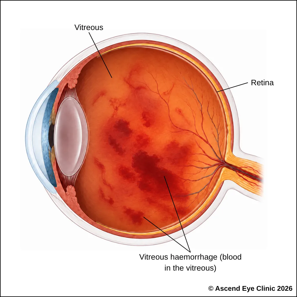


Symptoms usually develop suddenly and without pain. Vision may become hazy, smoky or cloudy, sometimes with a reddish or blood-stained tint. Some people notice shadows or missing areas in their vision, together with a sudden increase in floaters such as small dots, cobwebs, streaks or dark strands.
Flashes of light, a spreading shadow or a curtain across the vision indicate associated traction, a retinal tear or retinal detachment. These symptoms require urgent examination.
We use a combination of detailed eye examination and advanced retinal imaging to assess the macula, retina, optic nerve, and retinal blood vessels. These tests allow us to identify structural and functional changes, determine the type and severity of the condition, and establish a clear baseline for ongoing monitoring and treatment planning.
Measures how clearly you can see and helps identify any reduction in central vision.
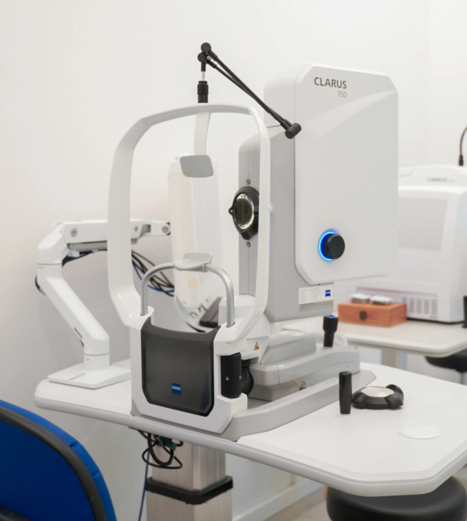
Captures images of the retina which gives an indication to the underlying cause of the vitreous hemorrhage. Widefield imaging allows us to capture the peripheral areas of the retina.
Provides cross-sectional images of the retina and macula, where the view allows, to identify retinal swelling, traction or other underlying changes associated with the bleeding.
Uses ultrasound waves to detect blood and other opacities within the vitreous cavity, while assessing the retina for associated retinal tears or detachment when the view is obscured by vitreous haemorrhage.
A small, stable vitreous haemorrhage can sometimes be observed while the blood is gradually absorbed by the eye. Vision often improves progressively over several weeks or months as the haemorrhage clears. Regular examinations remain important during this period, as the blood can obscure the view of the retina and hide an underlying retinal tear, retinal detachment or other retinal abnormality.
Further treatment depends on the underlying cause of the vitreous haemorrhage and whether the bleeding clears adequately. Treatment options include:
From your initial consultation to treatment and follow-up care, our team will guide you through every stage of your journey. We will take the time to explain your condition, answer your questions and help you feel informed and supported throughout your care.

Your consultation begins with a detailed assessment of your vision and eye health. Depending on your condition, this may include measurements of your vision and eye pressure, examination of the front and back of the eye, and advanced diagnostic imaging.
Our team will guide you through each test and explain what is being assessed. Your results will then be carefully reviewed to understand your individual needs and determine whether monitoring or treatment may be recommended.
Every eye and every patient is different. Your treatment plan will be tailored according to your examination findings, visual needs, general health and lifestyle.
We will explain the recommended treatment, its expected benefits, possible risks and any suitable alternatives in a clear and straightforward manner. You will have time to ask questions and consider your options before deciding how you would like to proceed.
Before your treatment, our team will guide you through the necessary preparations, paperwork and medication instructions. Where applicable, we will explain how to use your prescribed eye drops and how to care for your eyes afterwards.
We understand that undergoing a procedure can feel unfamiliar or worrying. Our team will be available to answer your questions and ensure that you know what to expect before the day of your treatment.
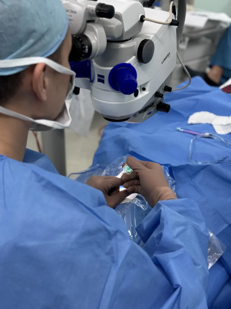
On the day of your procedure, you will arrive at the hospital or surgical centre where your operation has been scheduled. The surgical team will help you prepare and explain what will happen at each stage.
Anaesthetic eye drops or other forms of anaesthesia may be used, depending on the procedure, to keep you comfortable. Your doctor and the surgical team will support and reassure you throughout the operation.
Most eye surgeries are performed as day procedures, although the duration and recovery process will depend on the treatment you receive. Your vision may be temporarily blurred afterwards, so you should arrange for someone to accompany you home and should not drive unless your doctor has advised that it is safe to do so.
We will provide clear instructions on how to care for your eyes, use any prescribed medication and manage your activities during recovery. We will also explain which symptoms are expected and when you should contact the clinic for further advice.
Follow-up appointments allow us to monitor your vision, assess your recovery and identify any concerns at an early stage. Our team remains accessible should you have any questions or concerns throughout your recovery.
A vitreous haemorrhage can be caused by a retinal tear, retinal detachment or abnormal retinal blood vessels. Prompt assessment allows the source of bleeding to be identified and the appropriate treatment to begin.
Book a consultation at Ascend Eye Clinic for a detailed retinal examination.
Vitreous haemorrhage itself is usually painless. Patients often notice a sudden increase in floaters, hazy or smoky vision, a reddish tint to vision, or a marked reduction in sight without any eye pain.
Vitreous haemorrhage occurs when a blood vessel inside or around the retina bleeds into the vitreous, the clear gel filling the eye. Common causes include diabetic retinopathy, retinal tears, retinal vein occlusion, trauma and abnormal new blood vessel growth.
A sudden vitreous haemorrhage requires prompt assessment because the bleeding can sometimes be associated with a retinal tear or retinal detachment. We will examine the retina and determine whether urgent treatment is required.
Not necessarily. Vitreous haemorrhage has several possible causes, but a retinal tear or detachment is an important cause that must be excluded, particularly when the bleeding begins suddenly with new floaters or flashes.
Blood cells and small clumps of blood suspended within the vitreous cast shadows onto the retina. These can appear as numerous dots, strands, cobwebs, streaks or larger dark areas moving across your vision.
Blood within the vitreous interferes with light travelling through the eye before it reaches the retina. Depending on the amount of bleeding, vision can appear hazy, smoky, darkened or have a reddish or brownish tint.
A dense vitreous haemorrhage can cause a very significant reduction in vision, sometimes leaving only the ability to see large shapes, hand movements or light. Vitreous haemorrhage alone does not usually cause permanent complete blindness, but the underlying retinal condition can threaten vision if left untreated.
In many cases, the blood is gradually absorbed by the eye over time. Whether observation is appropriate depends on the cause of the bleeding, the amount of blood present and whether there is an underlying retinal condition that requires treatment.
Small haemorrhages can begin clearing over several weeks, while denser bleeding can take several months to resolve. We will monitor the eye during this period to ensure the blood is clearing and that the underlying retinal condition remains stable.
Blood within the vitreous can move and settle as you change position, so the amount of blood obscuring your central vision can vary throughout the day. This can make vision appear temporarily clearer or cloudier even while the overall haemorrhage is gradually resolving.
Persistent blood can continue to obstruct vision and prevent a clear view of the retina. If the haemorrhage remains dense or does not clear adequately, we can consider vitrectomy surgery to remove the blood-filled vitreous and directly assess and treat the retina.
If the haemorrhage is too dense to see through, we can use an ultrasound scan of the eye to assess the retina behind the blood and look for problems such as a retinal detachment. We will also repeat retinal examinations as the haemorrhage clears to obtain a clearer view of the retina.
Treatment is required when there is an underlying retinal condition that needs to be addressed, when bleeding continues or recurs, or when a dense haemorrhage does not clear sufficiently. Depending on the cause, treatment can include retinal laser, intravitreal injections or vitrectomy surgery.
The blood itself can clear without permanently damaging vision. The final visual outcome depends mainly on the condition that caused the bleeding and whether the retina or macula has been affected.
Vision can recover very well once the blood clears if the retina and macula remain healthy. If the haemorrhage resulted from a condition that has already affected the retina, the amount of visual recovery will depend on the extent of that retinal damage.
Yes. Recurrent bleeding can occur if the underlying source of the haemorrhage remains active, particularly with abnormal retinal blood vessels. Treating the underlying cause and continuing regular retinal follow-up help reduce the risk of further episodes.
We generally advise avoiding strenuous exercise and heavy lifting while an active vitreous haemorrhage is being assessed, particularly if there is concern about ongoing bleeding. We will advise you when it is appropriate to resume your usual activities based on the cause and stability of the retina.
Using a phone, computer or television does not worsen the haemorrhage. You can continue these activities as your vision allows, although tasks requiring fine detail can be difficult while the blood is obscuring your sight.
You should not drive if the haemorrhage has reduced your vision enough to affect your ability to see clearly and safely. We will assess your vision and advise when it is appropriate to return to driving.
Yes. Further bleeding can occur after the initial episode, causing a sudden increase in floaters, darker vision or a denser haze. Any sudden deterioration in vision should be assessed promptly so we can determine whether there is new bleeding or another retinal problem.
Hi, I would like to book a consultation with Dr Errol Chan from Ascend Eye Clinic
