
Keratoconus changes the shape and strength of the cornea over time. Early assessment and appropriate treatment can help stabilise the cornea, improve visual quality and protect vision for the future.
Keratoconus is a progressive eye condition in which the cornea gradually becomes thinner and develops an irregular, cone-like shape. The cornea is the clear front surface of the eye that focuses light onto the retina. When its shape becomes uneven, light no longer focuses clearly, resulting in blurred or distorted vision and irregular astigmatism.
In the early stages, keratoconus often appears as increasing short-sightedness or astigmatism, leading to frequent changes in spectacle prescription. As the cornea becomes more irregular, glasses and standard soft contact lenses provide less effective vision correction. Advanced keratoconus causes significant visual distortion, thinning and scarring of the cornea.
Early diagnosis allows the condition to be monitored closely and treated before further progression affects the corneal shape and vision.

Keratoconus often causes subtle visual changes in its early stages. As the condition progresses, symptoms become more noticeable and include blurred, distorted or double vision. Patients often experience increasing difficulty seeing clearly at night, with glare, halos and streaks around lights. Frequent changes in spectacle prescription and increasing astigmatism are also common as the cornea becomes progressively more irregular.


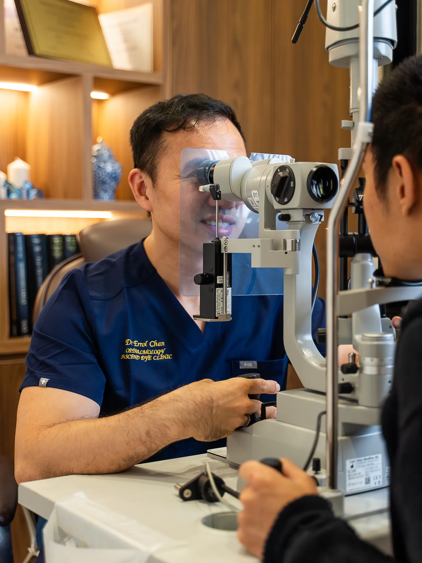
A detailed eye examination helps assess the shape, thickness and stability of the cornea, determine the severity of keratoconus and identify any progression over time. We will assess the cornea together with your vision and overall eye health to guide monitoring and treatment.
Measures how clearly you can see and helps identify any reduction in vision caused by irregular corneal shape.
Measures the eye’s focusing power and identifies changes in spectacle prescription and astigmatism caused by the irregular shape of the cornea.
Provides a magnified view of the cornea to identify structural changes associated with keratoconus, including corneal thinning, steepening and scarring.
Measures corneal thickness to identify areas of thinning and support assessment of disease severity and treatment planning.
Creates a detailed map of the corneal shape and curvature to detect irregular steepening, assess the extent of keratoconus and monitor changes over time.
Provides detailed cross-sectional images of the cornea to assess its thickness, shape and structural changes associated with keratoconus.
Treatment depends on the severity of keratoconus, whether the cornea is continuing to change and how much vision is affected. Early or stable keratoconus can be monitored with regular corneal imaging. Glasses or contact lenses can improve vision, while progressive keratoconus is treated with corneal collagen cross-linking to strengthen the cornea and reduce further progression. In more advanced cases of keratoconus, where the cornea has become severely thinned or scarred, corneal transplantation may be required to restore vision.
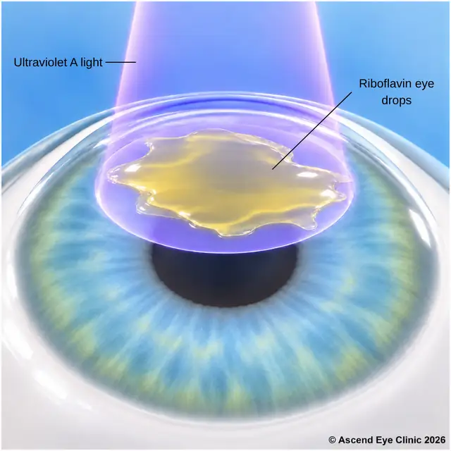
Corneal collagen cross-linking strengthens the cornea by increasing the bonds between collagen fibres within the corneal tissue. The aim is to stabilise the cornea and reduce further thinning and steepening caused by keratoconus.
Cross-linking is performed as an outpatient procedure under anaesthetic eye drops. The surface layer of the cornea is removed, allowing applied riboflavin eye drops to penetrate the corneal tissue. The cornea is then exposed to a controlled ultraviolet-A light, which activates the riboflavin and strengthens the collagen structure within the cornea. A bandage contact lens is placed over the eye at the end of the procedure while the surface heals.
The eye can feel sore, watery and sensitive to light during the first few days while the corneal surface recovers. Antibiotic and anti-inflammatory eye drops are prescribed, and the bandage contact lens is removed once healing is complete. Vision can fluctuate during the initial recovery period before gradually settling. Regular follow-up with corneal imaging is arranged to monitor healing and confirm that the keratoconus remains stable.
From your initial consultation to treatment and follow-up care, our team will guide you through every stage of your journey. We will take the time to explain your condition, answer your questions and help you feel informed and supported throughout your care.

Your consultation begins with a detailed assessment of your vision and eye health. Depending on your condition, this may include measurements of your vision and eye pressure, examination of the front and back of the eye, and advanced diagnostic imaging.
Our team will guide you through each test and explain what is being assessed. Your results will then be carefully reviewed to understand your individual needs and determine whether monitoring or treatment may be recommended.
Every eye and every patient is different. Your treatment plan will be tailored according to your examination findings, visual needs, general health and lifestyle.
We will explain the recommended treatment, its expected benefits, possible risks and any suitable alternatives in a clear and straightforward manner. You will have time to ask questions and consider your options before deciding how you would like to proceed.
Before your treatment, our team will guide you through the necessary preparations, paperwork and medication instructions. Where applicable, we will explain how to use your prescribed eye drops and how to care for your eyes afterwards.
We understand that undergoing a procedure can feel unfamiliar or worrying. Our team will be available to answer your questions and ensure that you know what to expect before the day of your treatment.
On the day of your procedure, you will arrive at the hospital or surgical centre where your operation has been scheduled. The surgical team will help you prepare and explain what will happen at each stage.
Anaesthetic eye drops or other forms of anaesthesia may be used, depending on the procedure, to keep you comfortable. Your doctor and the surgical team will support and reassure you throughout the operation.
Most eye surgeries are performed as day procedures, although the duration and recovery process will depend on the treatment you receive. Your vision may be temporarily blurred afterwards, so you should arrange for someone to accompany you home and should not drive unless your doctor has advised that it is safe to do so.
We will provide clear instructions on how to care for your eyes, use any prescribed medication and manage your activities during recovery. We will also explain which symptoms are expected and when you should contact the clinic for further advice.
Follow-up appointments allow us to monitor your vision, assess your recovery and identify any concerns at an early stage. Our team remains accessible should you have any questions or concerns throughout your recovery.
Keratoconus can continue to change the shape of the cornea over time, affecting both vision and the strength of the cornea. Early assessment helps identify progression and determine whether treatment such as corneal collagen cross-linking is appropriate.
Book a consultation for a detailed corneal assessment and personalised treatment plan.
Keratoconus develops when the cornea becomes thinner and gradually changes from a rounded shape into a more cone-like shape. Genetics, frequent eye rubbing and certain allergic or connective tissue conditions are associated with its development.
Keratoconus often progresses during the teenage years and early adulthood. Progression generally slows with age, but regular corneal imaging is important to detect changes in corneal shape and thickness.
As the cornea becomes steeper and more irregular, light no longer focuses evenly inside the eye. This causes blurred or distorted vision, increasing astigmatism, glare, halos and difficulty seeing clearly at night.
Yes. Keratoconus generally affects both eyes, although one eye is often more affected than the other.
Diagnosis involves examining the cornea and measuring its shape and thickness. Corneal topography or tomography creates detailed maps that detect characteristic changes and allow progression to be monitored over time.
Keratoconus alters the curvature of the cornea, causing increasing or irregular astigmatism. As the corneal shape changes, the spectacle prescription changes as well.
Glasses provide useful vision in early keratoconus when the corneal shape remains relatively regular. As irregularity increases, specialised contact lenses provide clearer vision by creating a smoother optical surface.
Options include rigid gas-permeable, hybrid and scleral contact lenses. The appropriate lens depends on the shape of the cornea, severity of keratoconus and the level of vision required.
Corneal collagen cross-linking strengthens the corneal tissue using riboflavin eye drops and controlled ultraviolet-A light. The treatment is used to stabilise progressive keratoconus and reduce further thinning and steepening.
The main purpose of cross-linking is to stabilise the cornea rather than remove the spectacle prescription. Vision continues to be corrected with glasses or contact lenses, although changes in corneal shape after treatment can alter vision over time.
Cross-linking is considered when examinations and corneal scans show that keratoconus is progressing. This includes changes in corneal curvature, thickness, vision or spectacle prescription.
Anaesthetic eye drops numb the eye during treatment. After the procedure, soreness, watering, light sensitivity and a gritty sensation are expected for the first few days while the surface of the cornea heals.
The surface of the cornea heals during the first several days. Vision remains blurred and fluctuates during the early recovery period before gradually stabilising over the following weeks to months.
Yes. Repeated or forceful eye rubbing places mechanical stress on the cornea and is associated with keratoconus progression. Allergies and persistent eye irritation should also be managed to reduce the urge to rub the eyes.
Most patients with keratoconus do not require corneal transplantation. Transplantation is considered in more severe cases when corneal scarring or irregularity prevents satisfactory vision despite glasses or specialised contact lenses.
Hi, I would like to book a consultation with Dr Errol Chan from Ascend Eye Clinic
